
12 months from date of receipt / reconstitution, -20 °C as supplied
| 应用 | 稀释度 |
|---|---|
| WB | 1:1000 |
| IHC-P | 1:250-1:1000 |
| ICC | 1:500 |
| ICFCM | 1:500 |
b-FGF, also known as FGF-2, is a potent mitogenic protein belonging to the fibroblast growth factor family. It plays a crucial role in cell proliferation, differentiation, and survival. b-FGF is particularly important for angiogenesis, wound healing, and tissue repair. It binds with high affinity to heparin and specifically to tyrosine kinase receptors, activating the FGF/FGFR signaling pathway, which in turn influences various cellular processes. b-FGF is widely distributed across different tissues, with highest concentrations found in the pituitary, brain, and neural tissues, retina, adrenal glands, and placenta. In cell culture, b-FGF is an essential growth factor for maintaining the undifferentiated state of stem cells, promoting the proliferation and differentiation of neural cells, and extending the survival of neurons. It also significantly promotes the proliferation and migration of endothelial cells when used in conjunction with heparin. The broad biological activities of b-FGF make it a key factor in biomedical research and clinical applications, particularly in regenerative medicine and the development of therapeutic strategies for various diseases.
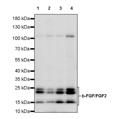
WB result of b-FGF/FGF2 Recombinant Rabbit mAb
Primary antibody: b-FGF/FGF2 Recombinant Rabbit mAb at 1/1000 dilution
Lane 1: HeLa whole cell lysate 20 µg
Lane 2: K562 whole cell lysate 20 µg
Lane 3: U-87 MG whole cell lysate 20 µg
Lane 4: SK-OV-3 whole cell lysate 20 µg
Secondary antibody: Goat Anti-rabbit IgG, (H+L), HRP conjugated at 1/10000 dilution
Predicted MW: 31 kDa
Observed MW: 15, 22, 24 kDa
Flow cytometric analysis of 4% PFA fixed 90% methanol permeabilized SK-OV-3 (Human ovarian cancer epithelial cell) labelling b-FGF/FGF2 antibody at 1/500 dilution (0.1 μg)/ (Red) compared with a Rabbit monoclonal IgG (Black) isotype control and an unlabelled control (cells without incubation with primary antibody and secondary antibody) (Blue). Goat Anti - Rabbit IgG Alexa Fluor® 488 was used as the secondary antibody.
IHC shows positive staining in paraffin-embedded human cerebral cortex. Anti- b-FGF/FGF2 antibody was used at 1/1000 dilution, followed by a HRP Polymer for Mouse & Rabbit IgG (ready to use). Counterstained with hematoxylin. Heat mediated antigen retrieval with Tris/EDTA buffer pH9.0 was performed before commencing with IHC staining protocol.
IHC shows positive staining in paraffin-embedded human kidney. Anti- b-FGF/FGF2 antibody was used at 1/1000 dilution, followed by a HRP Polymer for Mouse & Rabbit IgG (ready to use). Counterstained with hematoxylin. Heat mediated antigen retrieval with Tris/EDTA buffer pH9.0 was performed before commencing with IHC staining protocol.
IHC shows positive staining in paraffin-embedded human placenta. Anti- b-FGF/FGF2 antibody was used at 1/250 dilution, followed by a HRP Polymer for Mouse & Rabbit IgG (ready to use). Counterstained with hematoxylin. Heat mediated antigen retrieval with Tris/EDTA buffer pH9.0 was performed before commencing with IHC staining protocol.
IHC shows positive staining in paraffin-embedded human stomach. Anti- b-FGF/FGF2 antibody was used at 1/1000 dilution, followed by a HRP Polymer for Mouse & Rabbit IgG (ready to use). Counterstained with hematoxylin. Heat mediated antigen retrieval with Tris/EDTA buffer pH9.0 was performed before commencing with IHC staining protocol.
IHC shows positive staining in paraffin-embedded human prostate. Anti- b-FGF/FGF2 antibody was used at 1/250 dilution, followed by a HRP Polymer for Mouse & Rabbit IgG (ready to use). Counterstained with hematoxylin. Heat mediated antigen retrieval with Tris/EDTA buffer pH9.0 was performed before commencing with IHC staining protocol.
IHC shows positive staining in paraffin-embedded human hepatocellular carcinoma. Anti- b-FGF/FGF2 antibody was used at 1/250 dilution, followed by a HRP Polymer for Mouse & Rabbit IgG (ready to use). Counterstained with hematoxylin. Heat mediated antigen retrieval with Tris/EDTA buffer pH9.0 was performed before commencing with IHC staining protocol.
IHC shows positive staining in paraffin-embedded human thyroid cancer. Anti- b-FGF/FGF2 antibody was used at 1/250 dilution, followed by a HRP Polymer for Mouse & Rabbit IgG (ready to use). Counterstained with hematoxylin. Heat mediated antigen retrieval with Tris/EDTA buffer pH9.0 was performed before commencing with IHC staining protocol.
ICC shows positive staining in SK-OV-3 cells. Anti- b-FGF/FGF2 antibody was used at 1/500 dilution (Green) and incubated overnight at 4°C. Goat polyclonal Antibody to Rabbit IgG - H&L (Alexa Fluor® 488) was used as secondary antibody at 1/1000 dilution. The cells were fixed with 4% PFA and permeabilized with 0.1% PBS-Triton X-100. Nuclei were counterstained with DAPI (Blue). Counterstain with tubulin (Red).
 您现在的位置: