
PBS, 40% Glycerol, 0.05% BSA, 0.03% Proclin 300
12 months from date of receipt / reconstitution, -20 °C as supplied
| 应用 | 稀释度 |
|---|---|
| WB | 1:1000 |
| IP | 1:50 |
Phospho-TAK1 (Ser439) is a significant biomarker for the activation of TAK1 (Transforming growth factor-β activated kinase 1), which is a mitogen-activated protein kinase kinase kinase (MAP3K) involved in various cellular signaling pathways, including the MAPK and NF-κB pathways. The phosphorylation of TAK1 at serine 439 (or 412 in rodents) is a key event in its activation process. This modification allows TAK1 to interact with other proteins and initiate downstream signaling cascades that regulate cell growth, survival, and inflammation. The detection of Phospho-TAK1 (Ser439) is typically performed using specific antibodies that recognize this phosphorylated form of the protein.
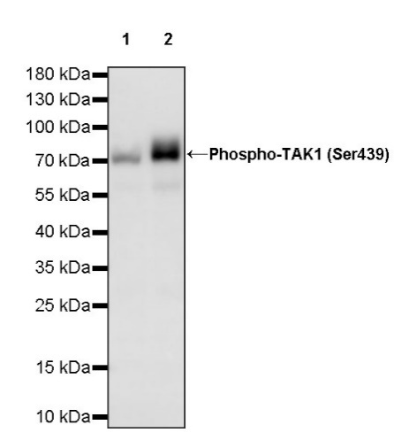
WB result of Phospho-TAK1 (Ser439) Recombinant Rabbit mAb
Primary antibody: Phospho-TAK1 (Ser439) Recombinant Rabbit mAb at 1/1000 dilution
Lane 1: untreated HeLa whole cell lysate 20 µg
Lane 2: HeLa treated with 100 nM Calyculin A and 20 ng/ml human IL-1β for 10 minutes whole cell lysate 20 µg
Secondary antibody: Goat Anti-rabbit IgG, (H+L), HRP conjugated at 1/10000 dilution
Predicted MW: 67 kDa
Observed MW: 75 kDa
Phospho-TAK1 (Ser439) Rabbit mAb at 1/50 dilution (1 µg) immunoprecipitating Phospho-TAK1 (Ser439) in 0.4 mg HeLa treated with 100 nM Calyculin A and 20 ng/ml human IL-1β for 10 minutes whole cell lysate.
Western blot was performed on the immunoprecipitate using Phospho-TAK1 (Ser439) Rabbit mAb at 1/1000 dilution.
Secondary antibody (HRP) for IP was used at 1/1000 dilution.
Lane 1: HeLa treated with 100 nM Calyculin A and 20 ng/ml human IL-1β for 10 minutes whole cell lysate 20 µg (Input)
Lane 2: Phospho-TAK1 (Ser439) Rabbit mAb IP in HeLa treated with 100 nM Calyculin A and 20 ng/ml human IL-1β for 10 minutes whole cell lysate
Lane 3: Rabbit monoclonal IgG IP in HeLa treated with 100 nM Calyculin A and 20 ng/ml human IL-1β for 10 minutes whole cell lysate
Predicted MW: 67 kDa
Observed MW: 75 kDa
 您现在的位置: