
12 months from date of receipt / reconstitution, -20 °C as supplied
| 应用 | 稀释度 |
|---|---|
| WB | 1:1000 |
| ICC | 1:200 |
PI3 Kinase p110 β, also known as PIK3CB, is a catalytic subunit of the phosphoinositide 3-kinase (PI3K) family, which plays a critical role in cellular processes such as growth, metabolism, and survival. PI3Ks are enzymes that generate phosphoinositol lipids as second messengers, initiating multiple signal transduction cascades. The p110 β isoform has been shown to have distinct functions in different cellular contexts and contributes to cell growth and development, DNA replication, insulin sensitivity, tumorigenesis, and G-protein-coupled receptor-activated signaling.
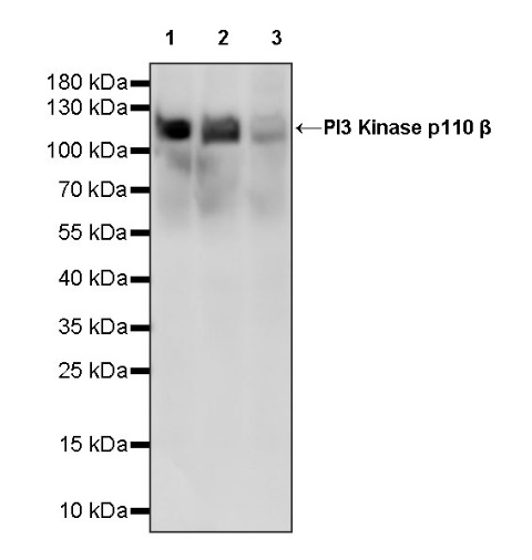
WB result of PI3 Kinase p110 β Recombinant Rabbit mAb
Primary antibody: PI3 Kinase p110 β Recombinant Rabbit mAb at 1/1000 dilution
Lane 1: K562 whole cell lysate 40 µg
Lane 2: MCF7 whole cell lysate 40 µg
Lane 3: HeLa whole cell lysate 40 µg
Secondary antibody: Goat Anti-rabbit IgG, (H+L), HRP conjugated at 1/10000 dilution
Predicted MW: 123 kDa
Observed MW: 100~130 kDa
WB result of PI3 Kinase p110 β Recombinant Rabbit mAb
Primary antibody: PI3 Kinase p110 β Recombinant Rabbit mAb at 1/1000 dilution
Lane 1: C2C12 whole cell lysate 40 µg
Lane 2: mouse brain lysate 40 µg
Secondary antibody: Goat Anti-rabbit IgG, (H+L), HRP conjugated at 1/10000 dilution
Predicted MW: 123 kDa
Observed MW: 110~150 kDa
WB result of PI3 Kinase p110 β Recombinant Rabbit mAb
Primary antibody: PI3 Kinase p110 β Recombinant Rabbit mAb at 1/1000 dilution
Lane 1: C6 whole cell lysate 40 µg
Lane 2: rat brain lysate 40 µg
Secondary antibody: Goat Anti-rabbit IgG, (H+L), HRP conjugated at 1/10000 dilution
Predicted MW: 123 kDa
Observed MW: 110~150 kDa
ICC shows positive staining in C2C12 cells. Anti- PI3 Kinase p110 β antibody was used at 1/200 dilution (Green) and incubated overnight at 4°C. Goat polyclonal Antibody to Rabbit IgG - H&L (Alexa Fluor® 488) was used as secondary antibody at 1/1000 dilution. The cells were fixed with 4% PFA and permeabilized with 0.1% PBS-Triton X-100. Nuclei were counterstained with DAPI (Blue). Counterstain with tubulin (Red).
ICC shows positive staining in HeLa cells. Anti- PI3 Kinase p110 β antibody was used at 1/200 dilution (Green) and incubated overnight at 4°C. Goat polyclonal Antibody to Rabbit IgG - H&L (Alexa Fluor® 488) was used as secondary antibody at 1/1000 dilution. The cells were fixed with 4% PFA and permeabilized with 0.1% PBS-Triton X-100. Nuclei were counterstained with DAPI (Blue). Counterstain with tubulin (Red).
 您现在的位置: