
PBS, 40% Glycerol, 0.05% BSA, 0.03% Proclin 300
12 months from date of receipt / reconstitution, -20 °C as supplied
| 应用 | 稀释度 |
|---|---|
| WB | 1:1000 |
| IHC-P | 1:500 |
| ICC | 1:500 |
| ICFCM | 1:50 |
The Stimulator of Interferon Genes (STING) protein is an integral component of the innate immune system, known for its role in mediating immune signaling pathways. Under normal conditions, STING resides in the endoplasmic reticulum (ER), and its activation is initiated through the cGAS-STING signaling pathway. Upon activation, STING is trafficked to the Golgi apparatus, which sets off a series of downstream signaling events. STING's journey from the ER to the Golgi apparatus is vital for its activation of downstream signaling pathways. Post-translational modifications, such as palmitoylation and phosphorylation, play a significant role in the trafficking and function of STING. The STING protein is also subject to regulation through ubiquitination, which is involved in its trafficking between the ER and Golgi apparatus. The E3 ubiquitin ligase RNF144 facilitates the K6-linked ubiquitination of STING, enhancing its transport. Furthermore, research has indicated that STING is involved in various cellular processes beyond the immune response, including autophagy, ER stress response, and metabolic reprogramming. After activation, STING is transported into small extracellular vesicles, where it can be recognized by the STAM protein, leading to the termination of STING signaling.
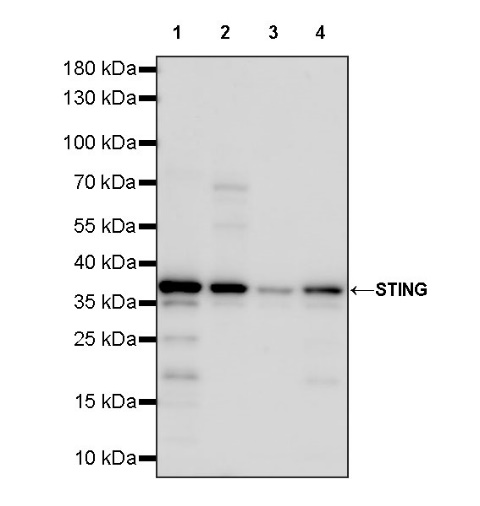
WB result of STING Recombinant Rabbit mAb
Primary antibody: STING Recombinant Rabbit mAb at 1/1000 dilution
Lane 1: THP-1 whole cell lysate 20 µg
Lane 2: HaCaT whole cell lysate 20 µg
Lane 3: HT-29 whole cell lysate 20 µg
Lane 4: HL-60 whole cell lysate 20 µg
Secondary antibody: Goat Anti-rabbit IgG, (H+L), HRP conjugated at 1/10000 dilution
Predicted MW: 42 kDa
Observed MW: 37 kDa
WB result of STING Recombinant Rabbit mAb
Primary antibody: STING Recombinant Rabbit mAb at 1/1000 dilution
Lane 1: A20 whole cell lysate 20 µg
Lane 2: RAW264.7 whole cell lysate 20 µg
Lane 3: NIH/3T3 whole cell lysate 20 µg
Lane 4: C2C12 whole cell lysate 20 µg
Secondary antibody: Goat Anti-rabbit IgG, (H+L), HRP conjugated at 1/10000 dilution
Predicted MW: 42 kDa
Observed MW: 37 kDa
Flow cytometric analysis of 4% PFA fixed 90% methanol permeabilized A549 (Human lung carcinoma epithelial cell, left) / THP-1 (Human monocytic leukemia monocyte, right) labelling STING antibody at 1/50 dilution (1 μg) / (Red) compared with a Rabbit monoclonal IgG (Black) isotype control and an unlabelled control (cells without incubation with primary antibody and secondary antibody) (Blue). Negative control: A549
IHC shows positive staining in paraffin-embedded human tonsil. Anti-STING antibody was used at 1/500 dilution, followed by a HRP Polymer for Mouse & Rabbit IgG (ready to use). Counterstained with hematoxylin. Heat mediated antigen retrieval with Tris/EDTA buffer pH9.0 was performed before commencing with IHC staining protocol.
IHC shows positive staining in paraffin-embedded human spleen. Anti-STING antibody was used at 1/500 dilution, followed by a HRP Polymer for Mouse & Rabbit IgG (ready to use). Counterstained with hematoxylin. Heat mediated antigen retrieval with Tris/EDTA buffer pH9.0 was performed before commencing with IHC staining protocol.
IHC shows positive staining in paraffin-embedded human lung. Anti-STING antibody was used at 1/500 dilution, followed by a HRP Polymer for Mouse & Rabbit IgG (ready to use). Counterstained with hematoxylin. Heat mediated antigen retrieval with Tris/EDTA buffer pH9.0 was performed before commencing with IHC staining protocol.
IHC shows positive staining in paraffin-embedded human endometrial cancer. Anti-STING antibody was used at 1/500 dilution, followed by a HRP Polymer for Mouse & Rabbit IgG (ready to use). Counterstained with hematoxylin. Heat mediated antigen retrieval with Tris/EDTA buffer pH9.0 was performed before commencing with IHC staining protocol.
IHC shows positive staining in paraffin-embedded human gastric cancer. Anti-STING antibody was used at 1/500 dilution, followed by a HRP Polymer for Mouse & Rabbit IgG (ready to use). Counterstained with hematoxylin. Heat mediated antigen retrieval with Tris/EDTA buffer pH9.0 was performed before commencing with IHC staining protocol.
IHC shows positive staining in paraffin-embedded mouse colon. Anti-STING antibody was used at 1/500 dilution, followed by a HRP Polymer for Mouse & Rabbit IgG (ready to use). Counterstained with hematoxylin. Heat mediated antigen retrieval with Tris/EDTA buffer pH9.0 was performed before commencing with IHC staining protocol.
ICC shows positive staining in THP-1 cells (top panel) and negative staining in A549 cells (below panel). Anti-STING antibody was used at 1/500 dilution (Green) and incubated overnight at 4°C. Goat polyclonal Antibody to Rabbit IgG - H&L (Alexa Fluor® 488) was used as secondary antibody at 1/1000 dilution. The cells were fixed with 4% PFA and permeabilized with 0.1% PBS-Triton X-100. Nuclei were counterstained with DAPI (Blue). Counterstain with tubulin (Red).
 您现在的位置: