
PBS, 40% Glycerol, 0.05% BSA, 0.03% Proclin 300
12 months from date of receipt / reconstitution, -20 °C as supplied
| 应用 | 稀释度 |
|---|---|
| WB | 1:1000 |
| IP | 1:50 |
| FCM | 1:500 |
CD5 is a cluster of differentiation expressed on the surface of T cells (various species) and in a subset of murine B cells known as B-1a. CD5 serves to mitigate activating signals from the BCR so that the B-1 cells can only be activated by very strong stimuli (such as bacterial proteins) and not by normal tissue proteins. There is no confirmed ligand for CD5 but there is evidence that CD72, a C-type lectin, may be a ligand or that CD5 may be homophilic, binding CD5 on the surface of other cells. T cells express higher levels of CD5 than B cells. CD5 is upregulated on T cells upon strong activation. In the thymus, there is a correlation with CD5 expression and strength of the interaction of the T cell towards self-peptides. CD5 is a good immunohistochemical marker for T-cells, although not as sensitive as CD3. About 76% of T-cell neoplasms are reported to express CD5, and it is also found in chronic lymphocytic leukemia and mantle cell lymphoma (both being B cell malignancies), that do not express CD3. It is commonly lost in cutaneous T-cell lymphoma, and its absence can be used as an indicator of malignancy in this condition. The absence of CD5 in T cell acute lymphoblastic leukemia, while relatively rare, is associated with a poor prognosis.
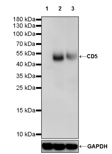
WB result of CD5 Rabbit pAb
Primary antibody: CD5 Rabbit pAb at 1/1000 dilution
Lane 1: Raji whole cell lysate 20 µg
Lane 2: Jurkat whole cell lysate 20 µg
Lane 3: Molt-4 whole cell lysate 20 µg
Negative control: Raji whole cell lysate
Secondary antibody: Goat Anti-rabbit IgG, (H+L), HRP conjugated at 1/10000 dilution
Predicted MW: 55 kDa
Observed MW: 50 kDa
Flow cytometric analysis of 4% PFA fixed 90% methanol permeabilized Raji (Human Burkitt's lymphoma B lymphocyte, left) / MOLT-4 (Human lymphoblastic leukemia T lymphoblast, right) labelling CD5 antibody at 1/500 dilution (0.1 μg) / (Red) compared with a Rabbit monoclonal IgG (Black) isotype control and an unlabelled control (cells without incubation with primary antibody and secondary antibody) (Blue). Goat Anti - Rabbit IgG Alexa Fluor® 488 was used as the secondary antibody.
Negative control: Raji
CD5 Rabbit pAb at 1/50 dilution (1 µg) immunoprecipitating CD5 in 0.4 mg Jurkat whole cell lysate.
Western blot was performed on the immunoprecipitate using CD5 Rabbit pAb at 1/1000 dilution.
Secondary antibody (HRP) for IP was used at 1/1000 dilution.
Lane 1: Jurkat whole cell lysate 20 µg (Input)
Lane 2: CD5 Rabbit pAb IP in Jurkat whole cell lysate
Lane 3: Rabbit monoclonal IgG IP in IP in IP in Jurkat whole cell lysate
Predicted MW: 55 kDa
Observed MW: 50 kDa
 您现在的位置: