
12 months from date of receipt / reconstitution, -20 °C as supplied
| 应用 | 稀释度 |
|---|---|
| WB | 1:1000 |
| IP | 1:50 |
| FCM | 1:50 |
CD58, also known as Lymphocyte Function-Associated Antigen 3 (LFA-3), is a glycoprotein that plays a critical role in immune cell interactions. It is widely expressed on various human tissue cells and acts as a counter-receptor for CD2, which is primarily found on the surface of T and NK cells. The interaction between CD2 and CD58 is an essential component of the immune synapse, facilitating not only cell adhesion but also inducing activation and proliferation of T/NK cells. This interaction triggers a series of intracellular signaling pathways in both the T/NK cells and target cells, which is crucial for immune responses. In addition to its membrane-bound form, a soluble form of CD58 (sCD58) exists in cell supernatants and tissues. sCD58 is considered an immunosuppressive factor that affects T/NK cell-mediated immune responses by influencing the CD2-CD58 interaction. Altered accumulation of sCD58 may contribute to immune suppression of T/NK cells in the tumor microenvironment, making sCD58 a potential target for immunotherapy.
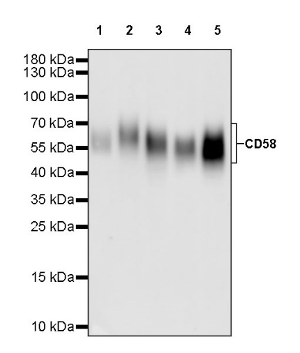
WB result of CD58 Recombinant Rabbit mAb
Primary antibody: CD58 Recombinant Rabbit mAb at 1/1000 dilution
Lane 1: Jurkat whole cell lysate 20 µg
Lane 2: K562 whole cell lysate 20 µg
Lane 3: Raji whole cell lysate 20 µg
Lane 4: SH-SY5Y whole cell lysate 20 µg
Lane 5: HeLa whole cell lysate 20 µg
Secondary antibody: Goat Anti-rabbit IgG, (H+L), HRP conjugated at 1/10000 dilution
Predicted MW: 28 kDa
Observed MW: 50~70 kDa
Flow cytometric analysis of human PBMC (human peripheral blood mononuclear cell) labelling CD58 antibody at 1/50 (1 μg) dilution (Right) compared with a Rabbit monoclonal IgG isotype control (Left). Goat Anti - Rabbit IgG Alexa Fluor® 488 was used as the secondary antibody. Then cells were stained with CD3 - Brilliant Violet 421™ separately. Gated on total viable cells.
CD58 Rabbit mAb at 1/50 dilution (1 µg) immunoprecipitating CD58 in 0.4 mg Raji whole cell lysate.
Western blot was performed on the immunoprecipitate using CD58 Rabbit mAb at 1/1000 dilution.
Secondary antibody (HRP) for IP was used at 1/1000 dilution.
Lane 1: Raji whole cell lysate 20 µg (Input)
Lane 2: CD58 Rabbit mAb IP in Raji whole cell lysate
Lane 3: Rabbit monoclonal IgG IP in Raji whole cell lysate
Predicted MW: 28 kDa
Observed MW: 50~70 kDa
 您现在的位置: