
PBS, 40% Glycerol, 0.05% BSA, 0.03% Proclin 300
12 months from date of receipt / reconstitution, -20 °C as supplied
| 应用 | 稀释度 |
|---|---|
| WB | 1:1000 |
| IP | 1:50 |
| ICC | 1:500 |
CXCL9, also known as MIG (monokine induced by interferon-gamma), is a member of the ELR-negative CXC chemokine subfamily that is inducible by IFN-γ. It plays a pivotal role in immune regulation and inflammation, and is implicated in various diseases, including cancer. CXCL9 is known to bind to its receptor CXCR3, facilitating the recruitment of CXCR3+ cells such as effector T cells, regulatory T cells (Tregs), and CD8+ cytotoxic T cells. This chemokine is involved in the pathogenesis of several physiological conditions, including its role in tumor growth, angiogenesis, and metastasis. In the context of cancer, CXCL9 has been identified as a potential biomarker and therapeutic target. It is suggested that the CXCL9/CXCR3 axis can be leveraged for cancer treatment by promoting the infiltration of immune cells to the tumor site, thereby enhancing anti-tumor immunity. Additionally, the expression levels of CXCL9 have been associated with the prognosis of certain cancers, indicating its potential as a prognostic indicator. Furthermore, research has highlighted the complex role of CXCL9 in the tumor microenvironment (TME). It can act as both an immunoactivator, promoting the recruitment of immune cells to combat tumor cells, and as a promoter of tumor growth and metastasis through autocrine signaling in certain contexts.
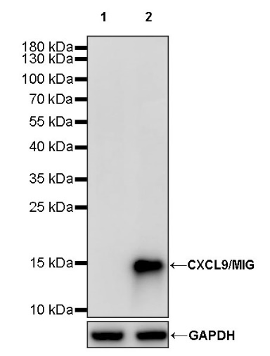
WB result of CXCL9/MIG Recombinant Rabbit mAb
Primary antibody: CXCL9/MIG Recombinant Rabbit mAb at 1/1000 dilution
Lane 1: untreated THP-1 whole cell lysate 20 µg
Lane 2: THP-1 treated with 200 ng/ml IFN-gamma and 50 ng/ml LPS for 24 hours whole cell lysate 20 µg
Secondary antibody: Goat Anti-rabbit IgG, (H+L), HRP conjugated at 1/10000 dilution
Predicted MW: 14 kDa
Observed MW: 15 kDa
CXCL9/MIG Rabbit mAb at 1/50 dilution (1 µg) immunoprecipitating CXCL9/MIG in 0.4 mg THP-1 treated with 200 ng/ml IFN-gamma and 50 ng/ml LPS for 24 hours whole cell lysate.
Western blot was performed on the immunoprecipitate using CXCL9/MIG Rabbit mAb at 1/1000 dilution.
Secondary antibody (HRP) for IP was used at 1/1000 dilution.
Lane 1: THP-1 treated with 200 ng/ml IFN-gamma and 50 ng/ml LPS for 24 hours whole cell lysate 20 µg (Input)
Lane 2: CXCL9/MIG Rabbit mAb IP in THP-1 treated with 200 ng/ml IFN-gamma and 50 ng/ml LPS for 24 hours whole cell lysate
Lane 3: Rabbit monoclonal IgG IP in THP-1 treated with 200 ng/ml IFN-gamma and 50 ng/ml LPS for 24 hours whole cell lysate
Predicted MW: 14 kDa
Observed MW: 15 kDa
ICC analysis of THP-1 cells treated with IFN-gamma(200ng/ml,24hr) and LPS (50 ng/ml, 24hr) (top panel) and untreated THP-1 cells (below panel). Anti-CXCL9/MIG antibody was used at 1/500 dilution (Green) and incubated overnight at 4°C. Goat polyclonal Antibody to Rabbit IgG - H&L (Alexa Fluor® 488) was used as secondary antibody at 1/1000 dilution. The cells were fixed with 4% PFA and permeabilized with 0.1% PBS-Triton X-100. Nuclei were counterstained with DAPI (Blue). Counterstain with tubulin (Red).
 您现在的位置: