
PBS, 40% Glycerol, 0.05% BSA, 0.03% Proclin 300
12 months from date of receipt / reconstitution, -20 °C as supplied
| 应用 | 稀释度 |
|---|---|
| WB | 1:1000 |
| IP | 1:50 |
| IHC-P | 1:1000 |
| ICC | 1:500 |
| ICFCM | 1:50 |
DYNLL1, also known as dynein light chain 1, is a protein that has been implicated in a variety of cellular processes, including DNA repair mechanisms. It is known to interact with the MRE11 protein, which is a component of the MRN complex that plays a key role in the initial steps of DNA double-strand break (DSB) repair. DYNLL1 is recruited to DSBs by 53BP1, where it modulates the extent of DNA end resection by binding to and destabilizing the MRE11 dimer. This interaction is crucial for the regulation of DNA repair pathway choice, particularly in the context of BRCA1-deficient cells, where it limits DNA end resection and thus influences homologous recombination. Furthermore, DYNLL1 has been identified as a component of the Shieldin complex, which is involved in the protection of DNA ends during replication stress and is implicated in the recruitment process of this complex to DSBs. In terms of its biological functions, DYNLL1 is essential for development and has been shown to promote endochondral bone formation by regulating intraflagellar dynein function in primary cilia. It is also involved in the regulation of the dynein motor complex, which is important for ciliary function and associated with ciliopathies, a group of disorders that affect the structure and function of cilia.
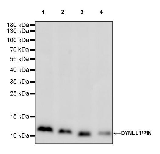
WB result of DYNLL1/PIN Recombinant Rabbit mAb
Primary antibody: DYNLL1/PIN Recombinant Rabbit mAb at 1/1000 dilution
Lane 1: 293T whole cell lysate 20 µg
Lane 2: HeLa whole cell lysate 20 µg
Lane 3: MCF7 whole cell lysate 20 µg
Lane 4: Jurkat whole cell lysate 20 µg
Secondary antibody: Goat Anti-rabbit IgG, (H+L), HRP conjugated at 1/10000 dilution
Predicted MW: 10 kDa
Observed MW: 11 kDa
WB result of DYNLL1/PIN Recombinant Rabbit mAb
Primary antibody: DYNLL1/PIN Recombinant Rabbit mAb at 1/1000 dilution
Lane 1: NIH/3T3 whole cell lysate 20 µg
Lane 2: mouse brain lysate 20 µg
Secondary antibody: Goat Anti-rabbit IgG, (H+L), HRP conjugated at 1/10000 dilution
Predicted MW: 10 kDa
Observed MW: 11 kDa
WB result of DYNLL1/PIN Recombinant Rabbit mAb
Primary antibody: DYNLL1/PIN Recombinant Rabbit mAb at 1/1000 dilution
Lane 1: C6 whole cell lysate 20 µg
Lane 2: rat brain lysate 20 µg
Secondary antibody: Goat Anti-rabbit IgG, (H+L), HRP conjugated at 1/10000 dilution
Predicted MW: 10 kDa
Observed MW: 11 kDa
Flow cytometric analysis of 4% PFA fixed 90% methanol permeabilized MCF7 (Human breast adenocarcinoma epithelial cell) labelling DYNLL1/PIN antibody at 1/50 dilution (1 μg)/ (Red) compared with a Rabbit monoclonal IgG (Black) isotype control and an unlabelled control (cells without incubation with primary antibody and secondary antibody) (Blue). Goat Anti - Rabbit IgG Alexa Fluor® 488 was used as the secondary antibody.
DYNLL1/PIN Rabbit mAb at 1/50 dilution (1 µg) immunoprecipitating DYNLL1/PIN in 0.4 mg HeLa whole cell lysate.
Western blot was performed on the immunoprecipitate using DYNLL1/PIN Rabbit mAb at 1/1000 dilution.
Secondary antibody (HRP) for IP was used at 1/1000 dilution.
Lane 1: HeLa whole cell lysate 20 µg (Input)
Lane 2: DYNLL1/PIN Rabbit mAb IP in HeLa whole cell lysate
Lane 3: Rabbit monoclonal IgG IP in HeLa whole cell lysate
Predicted MW: 10 kDa
Observed MW: 11 kDa
IHC shows positive staining in paraffin-embedded human cerebral cortex. Anti-DYNLL1/PIN antibody was used at 1/1000 dilution, followed by a HRP Polymer for Mouse & Rabbit IgG (ready to use). Counterstained with hematoxylin. Heat mediated antigen retrieval with Tris/EDTA buffer pH9.0 was performed before commencing with IHC staining protocol.
IHC shows positive staining in paraffin-embedded human lung squamous cell carcinoma. Anti-DYNLL1/PIN antibody was used at 1/1000 dilution, followed by a HRP Polymer for Mouse & Rabbit IgG (ready to use). Counterstained with hematoxylin. Heat mediated antigen retrieval with Tris/EDTA buffer pH9.0 was performed before commencing with IHC staining protocol.
IHC shows positive staining in paraffin-embedded mouse cerebral cortex. Anti-DYNLL1/PIN antibody was used at 1/1000 dilution, followed by a HRP Polymer for Mouse & Rabbit IgG (ready to use). Counterstained with hematoxylin. Heat mediated antigen retrieval with Tris/EDTA buffer pH9.0 was performed before commencing with IHC staining protocol.
IHC shows positive staining in paraffin-embedded rat cerebral cortex. Anti-DYNLL1/PIN antibody was used at 1/1000 dilution, followed by a HRP Polymer for Mouse & Rabbit IgG (ready to use). Counterstained with hematoxylin. Heat mediated antigen retrieval with Tris/EDTA buffer pH9.0 was performed before commencing with IHC staining protocol.
ICC shows positive staining in MCF7 cells. Anti-DYNLL1/PIN antibody was used at 1/500 dilution (Green) and incubated overnight at 4°C. Goat polyclonal Antibody to Rabbit IgG - H&L (Alexa Fluor® 488) was used as secondary antibody at 1/1000 dilution. The cells were fixed with 100% ice-cold methanol and permeabilized with 0.1% PBS-Triton X-100. Nuclei were counterstained with DAPI (Blue). Counterstain with tubulin (Red).
ICC shows positive staining in NIH/3T3 cells. Anti-DYNLL1/PIN antibody was used at 1/500 dilution (Green) and incubated overnight at 4°C. Goat polyclonal Antibody to Rabbit IgG - H&L (Alexa Fluor® 488) was used as secondary antibody at 1/1000 dilution. The cells were fixed with 4% PFA and permeabilized with 0.1% PBS-Triton X-100. Nuclei were counterstained with DAPI (Blue). Counterstain with tubulin (Red).
 您现在的位置: