
12 months from date of receipt / reconstitution, -20 °C as supplied
| 应用 | 稀释度 |
|---|---|
| WB | 1:4000 |
| IHC-P | 1:250 |
Insulin-degrading enzyme (IDE) is a key enzyme involved in the breakdown of insulin and has been implicated in the pathophysiology of both type 2 diabetes and Alzheimer's disease. IDE is capable of degrading a variety of substrates, including amyloid-β peptide, which is associated with Alzheimer's disease, as well as glucagon and islet amyloid polypeptide (IAPP), all of which have significant roles in glucose metabolism. The enzyme's function extends beyond its proteolytic role, as it also exhibits chaperone activity for amyloidogenic proteins and is involved in cellular proteostasis regulation. Recent research has highlighted the potential therapeutic applications of IDE in mitigating cognitive impairment in Alzheimer's disease and diabetes. The accumulation of amyloid-β in the brain is a common feature of these conditions, and IDE's ability to degrade these plaques makes it a promising target for treatment.
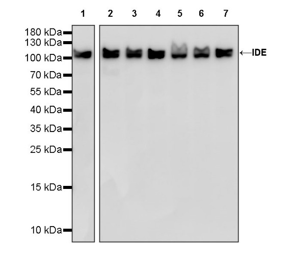
WB result of IDE Recombinant Rabbit mAb
Primary antibody: IDE Recombinant Rabbit mAb at 1/4000 dilution
Lane 1: Raji whole cell lysate 20 µg
Lane 2: HepG2 whole cell lysate 20 µg
Lane 3: A549 whole cell lysate 20 µg
Lane 4: K562 whole cell lysate 20 µg
Lane 5: HeLa whole cell lysate 20 µg
Lane 6: SK-BR-3 whole cell lysate 20 µg
Lane 7: MCF7 whole cell lysate 20 µg
Secondary antibody: Goat Anti-rabbit IgG, (H+L), HRP conjugated at 1/10000 dilution
Predicted MW: 118 kDa
Observed MW: 110 kDa
WB result of IDE Recombinant Rabbit mAb
Primary antibody: IDE Recombinant Rabbit mAb at 1/4000 dilution
Lane 1: mouse brain lysate 20 µg
Lane 2: mouse kidney lysate 20 µg
Lane 3: mouse liver lysate 20 µg
Secondary antibody: Goat Anti-rabbit IgG, (H+L), HRP conjugated at 1/10000 dilution
Predicted MW: 118 kDa
Observed MW: 110 kDa
WB result of IDE Recombinant Rabbit mAb
Primary antibody: IDE Recombinant Rabbit mAb at 1/4000 dilution
Lane 1: rat brain lysate 20 µg
Lane 2: rat kidney lysate 20 µg
Lane 3: rat liver lysate 20 µg
Secondary antibody: Goat Anti-rabbit IgG, (H+L), HRP conjugated at 1/10000 dilution
Predicted MW: 118 kDa
Observed MW: 110 kDa
IHC shows positive staining in paraffin-embedded human cervical squamous cell carcinoma. Anti-IDE antibody was used at 1/250 dilution, followed by a HRP Polymer for Mouse & Rabbit IgG (ready to use). Counterstained with hematoxylin. Heat mediated antigen retrieval with Tris/EDTA buffer pH9.0 was performed before commencing with IHC staining protocol.
IHC shows positive staining in paraffin-embedded human endometrial cancer. Anti-IDE antibody was used at 1/250 dilution, followed by a HRP Polymer for Mouse & Rabbit IgG (ready to use). Counterstained with hematoxylin. Heat mediated antigen retrieval with Tris/EDTA buffer pH9.0 was performed before commencing with IHC staining protocol.
IHC shows positive staining in paraffin-embedded human lung cancer. Anti-IDE antibody was used at 1/250 dilution, followed by a HRP Polymer for Mouse & Rabbit IgG (ready to use). Counterstained with hematoxylin. Heat mediated antigen retrieval with Tris/EDTA buffer pH9.0 was performed before commencing with IHC staining protocol.
IHC shows positive staining in paraffin-embedded human ovarian cancer. Anti-IDE antibody was used at 1/250 dilution, followed by a HRP Polymer for Mouse & Rabbit IgG (ready to use). Counterstained with hematoxylin. Heat mediated antigen retrieval with Tris/EDTA buffer pH9.0 was performed before commencing with IHC staining protocol.
IHC shows positive staining in paraffin-embedded human cerebral cortex. Anti-IDE antibody was used at 1/250 dilution, followed by a HRP Polymer for Mouse & Rabbit IgG (ready to use). Counterstained with hematoxylin. Heat mediated antigen retrieval with Tris/EDTA buffer pH9.0 was performed before commencing with IHC staining protocol.
IHC shows positive staining in paraffin-embedded human stomach. Anti-IDE antibody was used at 1/250 dilution, followed by a HRP Polymer for Mouse & Rabbit IgG (ready to use). Counterstained with hematoxylin. Heat mediated antigen retrieval with Tris/EDTA buffer pH9.0 was performed before commencing with IHC staining protocol.
 您现在的位置: