
PBS, 40% Glycerol, 0.05% BSA, 0.03% Proclin 300
12 months from date of receipt / reconstitution, -20 °C as supplied
| 应用 | 稀释度 |
|---|---|
| WB | 1:5000 |
| IHC-P | 1:1000 |
| ICC | 1:500 |
| ICFCM | 1:500 |
MCT4, or monocarboxylate transporter 4, is a plasmalemmal transporter that is instrumental in the release of lactate produced by astrocytes through aerobic glycolysis. It is characterized by its lower affinity for lactate compared to other monocarboxylate transporters, with a Km value of approximately 34 mM. MCT4 works in conjunction with MCT1 to facilitate the lactate shuttle, a process critical for the maintenance of lactate homeostasis in various tissues. MCT4 is particularly notable for its high expression in tumors and its role in maintaining intracellular pH homeostasis. Inhibiting MCT4 can suppress tumor growth by enhancing CD8+ T cell infiltration and cytotoxicity, which is linked to the alleviation of acidification in the tumor microenvironment and an increase in chemokine secretion induced by the reactive oxygen species/NF-κB signaling pathway. This suggests that MCT4 has a tumor-intrinsic function in creating an immunosuppressive environment in hepatocellular carcinoma (HCC), and targeting MCT4 could be a promising strategy to enhance the effectiveness of immunotherapy in HCC.
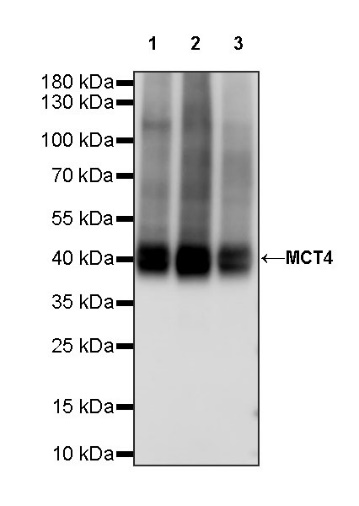
WB result of MCT4 Recombinant Rabbit mAb
Primary antibody: MCT4 Recombinant Rabbit mAb at 1/5000 dilution
Lane 1: unboiled HeLa whole cell lysate 20 µg
Lane 2: unboiled HepG2 whole cell lysate 20 µg
Lane 3: unboiled PC-3 whole cell lysate 20 µg
Secondary antibody: Goat Anti-rabbit IgG, (H+L), HRP conjugated at 1/10000 dilution
Predicted MW: 49 kDa
Observed MW: 45 kDa
Flow cytometric analysis of 4% PFA fixed 90% methanol permeabilized HeLa (Human cervix adenocarcinoma epithelial cell) labelling MCT4 antibody at 1/500 dilution (0.1 μg)/ (Red) compared with a Rabbit monoclonal IgG (Black) isotype control and an unlabelled control (cells without incubation with primary antibody and secondary antibody) (Blue). Goat Anti - Rabbit IgG Alexa Fluor® 488 was used as the secondary antibody.
IHC shows positive staining in paraffin-embedded human placenta. Anti-MCT4 antibody was used at 1/1000 dilution, followed by a HRP Polymer for Mouse & Rabbit IgG (ready to use). Counterstained with hematoxylin. Heat mediated antigen retrieval with Tris/EDTA buffer pH9.0 was performed before commencing with IHC staining protocol.
IHC shows positive staining in paraffin-embedded human testis. Anti-MCT4 antibody was used at 1/1000 dilution, followed by a HRP Polymer for Mouse & Rabbit IgG (ready to use). Counterstained with hematoxylin. Heat mediated antigen retrieval with Tris/EDTA buffer pH9.0 was performed before commencing with IHC staining protocol.
IHC shows positive staining in paraffin-embedded human lung. Anti-MCT4 antibody was used at 1/1000 dilution, followed by a HRP Polymer for Mouse & Rabbit IgG (ready to use). Counterstained with hematoxylin. Heat mediated antigen retrieval with Tris/EDTA buffer pH9.0 was performed before commencing with IHC staining protocol.
IHC shows positive staining in paraffin-embedded human colon cancer. Anti-MCT4 antibody was used at 1/1000 dilution, followed by a HRP Polymer for Mouse & Rabbit IgG (ready to use). Counterstained with hematoxylin. Heat mediated antigen retrieval with Tris/EDTA buffer pH9.0 was performed before commencing with IHC staining protocol.
IHC shows positive staining in paraffin-embedded human lung squamous cell carcinoma. Anti-MCT4 antibody was used at 1/1000 dilution, followed by a HRP Polymer for Mouse & Rabbit IgG (ready to use). Counterstained with hematoxylin. Heat mediated antigen retrieval with Tris/EDTA buffer pH9.0 was performed before commencing with IHC staining protocol.
IHC shows positive staining in paraffin-embedded mouse skeletal muscle. Anti-MCT4 antibody was used at 1/1000 dilution, followed by a HRP Polymer for Mouse & Rabbit IgG (ready to use). Counterstained with hematoxylin. Heat mediated antigen retrieval with Tris/EDTA buffer pH9.0 was performed before commencing with IHC staining protocol.
ICC shows positive staining in HeLa cells. Anti- MCT4 antibody was used at 1/500 dilution (Green) and incubated overnight at 4°C. Goat polyclonal Antibody to Rabbit IgG - H&L (Alexa Fluor® 488) was used as secondary antibody at 1/1000 dilution. The cells were fixed with 4% PFA and permeabilized with 0.1% PBS-Triton X-100. Nuclei were counterstained with DAPI (Blue). Counterstain with tubulin (Red).
 您现在的位置: