
12 months from date of receipt / reconstitution, -20 °C as supplied
| 应用 | 稀释度 |
|---|---|
| WB | 1:1000 |
| IP | 1:50 |
| IHC-P | 1:200 |
PAK1, or p21-activated kinase 1, is a serine/threonine kinase known for its role as an effector of the small GTPases Rac1 and Cdc42. It is involved in a plethora of cellular functions, including cytoskeletal dynamics, cell cycle regulation, cell survival, and apoptosis. Research has highlighted the significance of PAK1 in aging and longevity. In the nematode Caenorhabditis elegans, PAK1 function limits lifespan under basal conditions by inhibiting the forkhead box transcription factor DAF-16. Interestingly, the depletion of PAK extends lifespan and ameliorates age-related phenotypes in a premature aging mouse model, suggesting that PAK is not only an oncogenic kinase but also an aging kinase. Moreover, PAK1 has been identified as a potential therapeutic target in cancer due to its involvement in multiple oncogenic signaling pathways. The kinase also plays a central role in major cell proliferation pathways, such as the Wnt/β-catenin and EGFR/HER2/MAPK pathways, and is involved in the regulation of apoptosis and NF-κB pathways, cell cycle progression, and autophagy.
WB result of PAK1 Rabbit pAb
Primary antibody: PAK1 Rabbit pAb at 1/1000 dilution
Lane 1: SK-OV-3 whole cell lysate 20 µg
Lane 2: 293T whole cell lysate 20 µg
Lane 3: SH-SY5Y whole cell lysate 20 µg
Secondary antibody: Goat Anti-rabbit IgG, (H+L), HRP conjugated at 1/10000 dilution
Predicted MW: 61 kDa
Observed MW: 65 kDa
WB result of PAK1 Rabbit pAb
Primary antibody: PAK1 Rabbit pAb at 1/1000 dilution
Lane 1: NIH/3T3 whole cell lysate 20 µg
Secondary antibody: Goat Anti-rabbit IgG, (H+L), HRP conjugated at 1/10000 dilution
Predicted MW: 61 kDa
Observed MW: 65 kDa
WB result of PAK1 Rabbit pAb
Primary antibody: PAK1 Rabbit pAb at 1/1000 dilution
Lane 1: C6 whole cell lysate 20 µg
Secondary antibody: Goat Anti-rabbit IgG, (H+L), HRP conjugated at 1/10000 dilution
Predicted MW: 61 kDa
Observed MW: 65 kDa
PAK1 Rabbit pAb at 1/50 dilution (1 µg) immunoprecipitating PAK1 in 0.4 mg 293T whole cell lysate.
Western blot was performed on the immunoprecipitate using PAK1 Rabbit pAb at 1/1000 dilution.
Secondary antibody (HRP) for IP was used at 1/1000 dilution.
Lane 1: 293T whole cell lysate 20 µg (Input)
Lane 2: PAK1 Rabbit pAb IP in 293T whole cell lysate
Lane 3: Rabbit monoclonal IgG IP in 293T whole cell lysate
Predicted MW: 61 kDa
Observed MW: 65 kDa
IHC shows positive staining in paraffin-embedded human cerebral cortex. Anti-PAK1 antibody was used at 1/200 dilution, followed by a HRP Polymer for Mouse & Rabbit IgG (ready to use). Counterstained with hematoxylin. Heat mediated antigen retrieval with Tris/EDTA buffer pH9.0 was performed before commencing with IHC staining protocol.
IHC shows positive staining in paraffin-embedded human colon cancer. Anti-PAK1 antibody was used at 1/200 dilution, followed by a HRP Polymer for Mouse & Rabbit IgG (ready to use). Counterstained with hematoxylin. Heat mediated antigen retrieval with Tris/EDTA buffer pH9.0 was performed before commencing with IHC staining protocol.
IHC shows positive staining in paraffin-embedded mouse cerebral cortex. Anti-PAK1 antibody was used at 1/200 dilution, followed by a HRP Polymer for Mouse & Rabbit IgG (ready to use). Counterstained with hematoxylin. Heat mediated antigen retrieval with Tris/EDTA buffer pH9.0 was performed before commencing with IHC staining protocol.
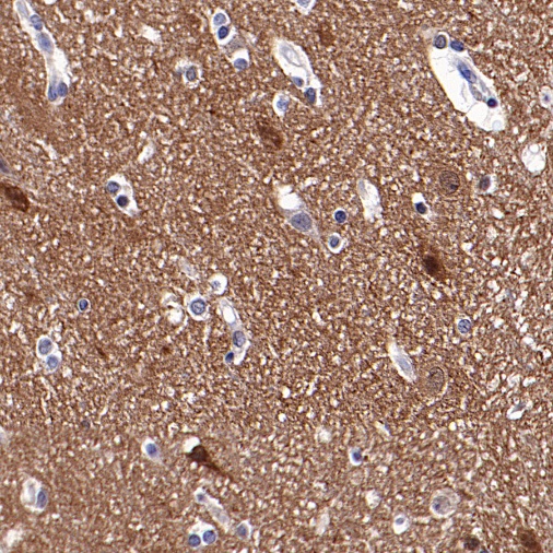
IHC shows positive staining in paraffin-embedded rat cerebral cortex. Anti-PAK1 antibody was used at 1/200 dilution, followed by a HRP Polymer for Mouse & Rabbit IgG (ready to use). Counterstained with hematoxylin. Heat mediated antigen retrieval with Tris/EDTA buffer pH9.0 was performed before commencing with IHC staining protocol.
 您现在的位置: