
PBS, 40% Glycerol, 0.05% BSA, 0.03% Proclin 300
12 months from date of receipt / reconstitution, -20 °C as supplied
| 应用 | 稀释度 |
|---|---|
| Dot Blot | 1:1000 |
| WB | 1:1000 |
| IP | 1:50 |
| ICC | 1:500 |
p38 mitogen-activated protein kinases are a class of mitogen-activated protein kinases (MAPKs) that are responsive to stress stimuli, such as cytokines, ultraviolet irradiation, heat shock, and osmotic shock, and are involved in cell differentiation, apoptosis and autophagy. Abnormal activity (higher or lower than physiological) of p38 has been implicated in pathological stresses in several tissues, that include neuronal, bone, lung, cardiac and skeletal muscle, red blood cells, and fetal tissues. Phospho-p38 MAPK (Tyr182), often tested alongside Thr180, is a significant biomarker for the activation state of p38 MAPK, a crucial enzyme in cellular stress and inflammatory responses. The phosphorylation at these sites, Thr180 and Tyr182, is essential for the full activation of p38 MAPK, allowing it to regulate various cellular processes in response to stress, inflammation, and other signals.
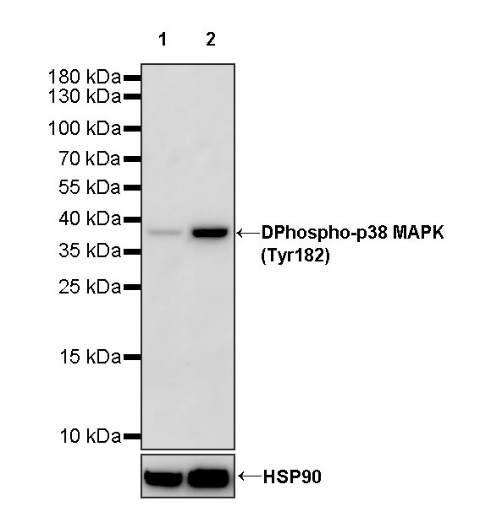
WB result of Phospho-p38 MAPK (Tyr182) Recombinant Rabbit mAb
Primary antibody: Phospho-p38 MAPK (Tyr182) Recombinant Rabbit mAb at 1/1000 dilution
Lane 1: untreated Jurkat whole cell lysate 20 µg
Lane 2: Jurkat treated with 25 μM Anisomycin for 30 minutes whole cell lysate 20 µg
Secondary antibody: Goat Anti-rabbit IgG, (H+L), HRP conjugated at 1/10000 dilution
Predicted MW: 41 kDa
Observed MW: 38 kDa
WB result of Phospho-p38 MAPK (Tyr182) Recombinant Rabbit mAb
Primary antibody: Phospho-p38 MAPK (Tyr182) Recombinant Rabbit mAb at 1/1000 dilution
Lane 1: untreated NIH/3T3 whole cell lysate 20 µg
Lane 2: NIH/3T3 treated with 25 μM Anisomycin for 30 minutes whole cell lysate 20 µg
Secondary antibody: Goat Anti-rabbit IgG, (H+L), HRP conjugated at 1/10000 dilution
Predicted MW: 41 kDa
Observed MW: 38 kDa
Dot blot result of Phospho-p38 MAPK (Tyr182) Recombinant Rabbit mAb
Lane 1: Phospho-p38 MAPK (Thr180/Tyr182) peptide
Lane 2: p38 MAPK WT peptide
Lane 3: Phospho-p38 MAPK (Thr180) peptide
Lane 4: Phospho-p38 MAPK (Tyr182) peptide
Primary antibody: Phospho-p38 MAPK (Tyr182) Recombinant Rabbit mAb at 1/1000 dilution
Secondary antibody: Goat Anti-rabbit IgG, (H+L), HRP conjugated at 1/10000 dilution
ICC analysis of Jurkat cells treated with Anisomycin (25μM, 30min) (top panel) and Jurkat cells untreated with Anisomycin (25μM, 30min) (below panel). Anti-Phospho-p38 MAPK (Tyr182) antibody was used at 1/500 dilution (Green) and incubated overnight at 4°C. Goat polyclonal Antibody to Rabbit IgG - H&L (Alexa Fluor® 488) was used as secondary antibody at 1/1000 dilution. The cells were fixed with 4% PFA and permeabilized with 0.1% PBS-Triton X-100. Nuclei were counterstained with DAPI (Blue). Counterstain with tubulin (Red).
 您现在的位置: