
PBS, 40% Glycerol, 0.05% BSA, 0.03% Proclin 300
12 months from date of receipt / reconstitution, -20 °C as supplied
| 应用 | 稀释度 |
|---|---|
| WB | 1:1000 |
| IP | 1:50 |
| IHC-P | 1:500 |
| IF | 1:200 |
TIM-3, also known as Hepatitis A virus Cell receptor 2 (HAVCR2), is a transmembrane protein and a member of the TIM protein family, which plays a crucial role in immune response regulation. It is expressed on various immune cells, including CD8+ T cells, Th1 cells, regulatory T cells (Tregs), dendritic cells (DCs), natural killer (NK) cells, monocytes, and macrophages. TIM-3 functions as an inhibitory receptor and is associated with T cell exhaustion in chronic infections and cancer. It has been identified as a promising target for cancer immunotherapy, as its blockade can enhance antitumor immunity. The receptor interacts with several ligands, including Galectin-9, CEACAM-1, phosphatidylserine (PtdSer), and High Mobility Group Box 1 (HMGB1). TIM-3 also plays a role in the clearance of apoptotic bodies by interacting with PtdSer on the surface of apoptotic cells. Recent studies have shown that targeting TIM-3 on DCs might be more critical for enhancing antitumor immunity than targeting it on T cells. Furthermore, combined blockade of TIM-3 and PD-1 has shown synergistic effects in reducing tumor growth in various mouse tumor models, suggesting that the combination therapy might be a promising approach for cancer treatment.
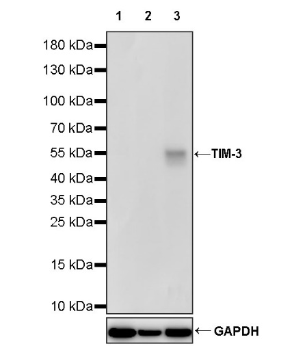
WB result of TIM-3 Recombinant Rabbit mAb
Primary antibody: TIM-3 Recombinant Rabbit mAb at 1/1000 dilution
Lane 1: Jurkat whole cell lysate 20 µg
Lane 2: HT-29 whole cell lysate 20 µg
Lane 3: Daudi whole cell lysate 20 µg
Negative control: Jurkat whole cell lysate; HT-29 whole cell lysate
Secondary antibody: Goat Anti-rabbit IgG, (H+L), HRP conjugated at 1/10000 dilution
Predicted MW: 33 kDa
Observed MW: 55 kDa
TIM-3 Rabbit mAb at 1/50 dilution (1 µg) immunoprecipitating TIM-3 in 0.4 mg Daudi whole cell lysate.
Western blot was performed on the immunoprecipitate using TIM-3 Rabbit mAb at 1/1000 dilution.
Secondary antibody (HRP) for IP was used at 1/1000 dilution.
Lane 1: Daudi whole cell lysate 20 µg (Input)
Lane 2: TIM-3 Rabbit mAb IP in Daudi whole cell lysate
Lane 3: Rabbit monoclonal IgG IP in Daudi whole cell lysate
Predicted MW: 33 kDa
Observed MW: 55 kDa
This blot was developed with high sensitivity substrate
IHC shows positive staining in paraffin-embedded human tonsil. Anti- TIM-3 antibody was used at 1/500 dilution, followed by a HRP Polymer for Mouse & Rabbit IgG (ready to use). Counterstained with hematoxylin. Heat mediated antigen retrieval with Tris/EDTA buffer pH9.0 was performed before commencing with IHC staining protocol.
IHC shows positive staining in paraffin-embedded human liver. Anti- TIM-3 antibody was used at 1/500 dilution, followed by a HRP Polymer for Mouse & Rabbit IgG (ready to use). Counterstained with hematoxylin. Heat mediated antigen retrieval with Tris/EDTA buffer pH9.0 was performed before commencing with IHC staining protocol.
IHC shows positive staining in paraffin-embedded human stomach. Anti- TIM-3 antibody was used at 1/500 dilution, followed by a HRP Polymer for Mouse & Rabbit IgG (ready to use). Counterstained with hematoxylin. Heat mediated antigen retrieval with Tris/EDTA buffer pH9.0 was performed before commencing with IHC staining protocol.
IHC shows positive staining in paraffin-embedded human hepatocellular carcinoma. Anti- TIM-3 antibody was used at 1/500 dilution, followed by a HRP Polymer for Mouse & Rabbit IgG (ready to use). Counterstained with hematoxylin. Heat mediated antigen retrieval with Tris/EDTA buffer pH9.0 was performed before commencing with IHC staining protocol.
IHC shows positive staining in paraffin-embedded human colon cancer. Anti- TIM-3 antibody was used at 1/500 dilution, followed by a HRP Polymer for Mouse & Rabbit IgG (ready to use). Counterstained with hematoxylin. Heat mediated antigen retrieval with Tris/EDTA buffer pH9.0 was performed before commencing with IHC staining protocol.
IHC shows positive staining in paraffin-embedded human endometrial cancer. Anti- TIM-3 antibody was used at 1/500 dilution, followed by a HRP Polymer for Mouse & Rabbit IgG (ready to use). Counterstained with hematoxylin. Heat mediated antigen retrieval with Tris/EDTA buffer pH9.0 was performed before commencing with IHC staining protocol.
IF shows positive staining in paraffin-embedded human tonsil. Anti-TIM-3 antibody was used at 1/200 dilution (Green) and incubated overnight at 4°C. Goat polyclonal Antibody to Rabbit IgG - H&L (Alexa Fluor® 488) was used as secondary antibody at 1/1000 dilution. Counterstained with DAPI (Blue). Heat mediated antigen retrieval with EDTA buffer pH9.0 was performed before commencing with IF staining protocol.
IF shows positive staining in paraffin-embedded human colon cancer. Anti-TIM-3 antibody was used at 1/200 dilution (Green) and incubated overnight at 4°C. Goat polyclonal Antibody to Rabbit IgG - H&L (Alexa Fluor® 488) was used as secondary antibody at 1/1000 dilution. Counterstained with DAPI (Blue). Heat mediated antigen retrieval with EDTA buffer pH9.0 was performed before commencing with IF staining protocol.
 您现在的位置: