
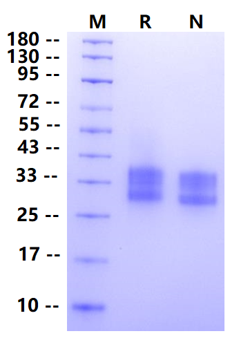
28-40kDa (Reducing)
Reconstitute at 0.1-1 mg/ml according to the size in ultrapure water after rapid centrifugation.
1. Sempowski G D. et al. (1999) Resistance of CD7-deficient mice to lipopolysaccharide-induced shock syndromes. J Exp Med. 189(6): 1011-1016.
CD7 is a 40-kD member of the Ig gene superfamily that is expressed on a major subset of human peripheral T lymphocytes and NK cells. CD7 is an early T cell activation antigen in that CD7 mRNA levels rise within 15 min after initiation of a transmembrane calcium ion flux. CD7 can complex with CD3 and CD45 molecules, and CD7 signaling involves both protein kinase C and protein tyrosine kinase. CD7 has been shown to be a functional signal-transducing molecule on resting NK cells. Antibody cross-linking of NK cell CD7 induced increases in free cytoplasmic calcium, secretion of IFN-γ, NK cell proliferation, adhesion to fibronectin, and NK cytotoxic activity. Although the above studies have demonstrated in vitro roles for CD7 in T and NK cell activation and/or adhesion, relevant functions of CD7 in vivo remain unknown.
2μg (R: reducing condition, N: non-reducing condition).
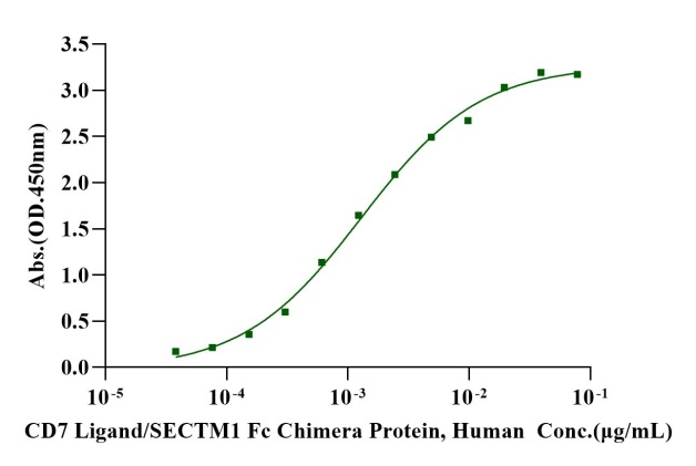
Immobilized Biotinylated CD7 His&Avi Tag Protein, Human (Cat. No. UA010725) at 2 μg/mL on Streptavidin precoated (0.5μg/well) plate, can bind CD7 Ligand/SECTM1 Fc Chimera, Human (Cat. No. UA010409) with EC50 of 1.03-1.61ng/ml.
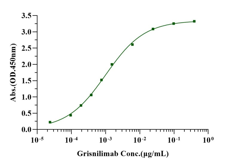
Immobilized Biotinylated CD7 His&Avi Tag, Human (Cat. No. UA010725) at 2 μg/mL on Streptavidin precoated (0.5μg/well) plate, can bind Grisnilimab (Cat. No. UA011033) with EC50 of 0.83-1.20 ng/ml.
 您现在的位置: