

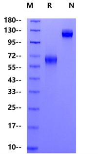
Leu25-Gln167 with C-terminal Human IgG1 Fc
LEVPNGPWRSLTFYPAWLTVSEGANATFTCSLSNWSEDLMLNWNRLSPSNQTEKQAAFCNGLSQPVQDARFQIIQLPNRHDFHMNILDTRRNDSGIYLCGAISLHPKAKIEESPGAELVVTERILETSTRYPSPSPKPEGRFQIEGRMDPKSSDKTHTCPPCPAPELLGGPSVFLFPPKPKDTLMISRTPEVTCVVVDVSHEDPEVKFNWYVDGVEVHNAKTKPREEQYNSTYRVVSVLTVLHQDWLNGKEYKCKVSNKALPAPIEKTISKAKGQPREPQVYTLPPSRDELTKNQVSLTCLVKGFYPSDIAVEWESNGQPENNYKTTPPVLDSDGSFFLYSKLTVDKSRWQQGNVFSCSVMHEALHNHYTQKSLSLSPGK
1、James E S. et al. (2005) PDCD1: a tissue-specific susceptibility locus for inherited inflammatory disorders. Genes Immun. 6(5): 430-437.
2、Okazaki T. et al. (2007) PD-1 and PD-1 ligands: from discovery to clinical application. Int Immunol. 19(7): 813-824.
3、del Rio M L. et al. (2008) PD-1/PD-L1, PD-1/PD-L2, and other co-inhibitory signaling pathways in transplantation. Transpl Int. 21(11): 1015-1028.
4、Riley J L. (2009) PD-1 signaling in primary T cells. Immunol Rev. 229(1): 114-25.
Programmed cell death protein 1, also known as PD-1 and CD279 (cluster of differentiation 279) or PDCD1, is a protein that in humans is encoded by the PDCD1 gene. Mature mouse PD-1 consists of a 149 amino acid (aa) extracellular region (ECD) with one immunoglobulin-like V-type domain, a 21 aa transmembrane domain, and a 98 aa cytoplasmic region. The mouse PD-1 ECD shares 65% aa sequence identity with the human PD-1 ECD. The cytoplasmic tail contains two tyrosine residues that form the immunoreceptor tyrosine-based inhibitory motif (ITIM) and immunoreceptor tyrosine-based switch motif (ITSM) that are important for mediating PD-1 signaling.
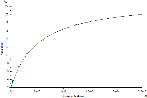
PD-1 Fc Chimera,Mouse (Cat. No. UA010222) captured on Protein A Biosenor, can bind PD-L1/B7-H1 His Tag, Mouse (Cat. No. UA010223) with an affinity constant of 0.49μM as determined in SPR assay.
 您现在的位置: