
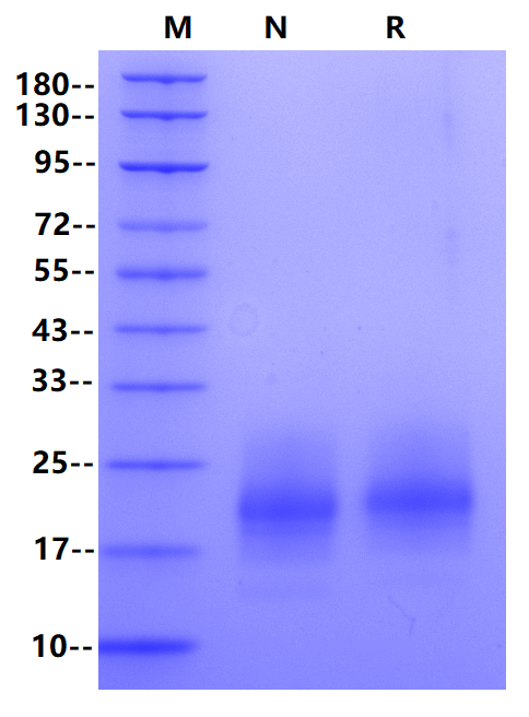
Ala18-Glu144
20-25 kDa(Reducing)
PBS, pH7.4
Reconstitute at less than 1 mg/mL according to the size in ultrapure water after rapid centrifugation .
· 12 months from date of receipt, lyophilized powder stored at -20 to -80℃.
· 3 months, -20 to -80℃ under sterile conditions after reconstitution.
· 1 week, 2 to 8℃ under sterile conditions after reconstitution.
· Please avoid repeated freeze-thaw cycles.
Granulocyte-macrophage colony-stimulating factor (GM-CSF), also known as colony-stimulating factor 2 (CSF2), is a monomeric glycoprotein secreted by macrophages, T cells, mast cells, natural killer cells, endothelial cells and fibroblasts that functions as a cytokine. GM-CSF was first described as a growth factor that induces the differentiation and proliferation of myeloid progenitors in the bone marrow, which also has an important cytokine effect in chronic inflammatory diseases by stimulating the activation and migration of myeloid cells to inflammation sites, promoting survival of target cells and stimulating the renewal of effector granulocytes and macrophages. GM-CSF receptor is composed of one α chain and one β chain with low and high-affinity binding to GM-CSF, respectively, and the β chain is shared with IL-3 and IL-5 receptor. GM-CSF signals via signal transducer and activator of transcription, STAT5. In macrophages, it has also been shown to signal via STAT3. The cytokine activates macrophages to inhibit fungal survival.
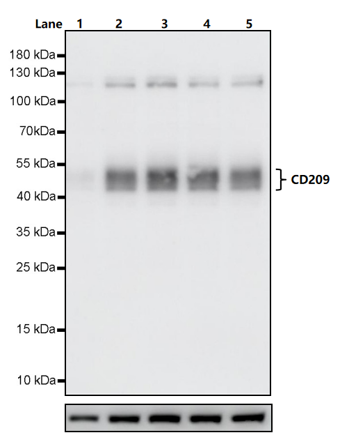
Primary antibody: CD209 gamma Recombinant Rabbit mAb (S0B1390) at 1/1000 dilution
Lane 1: untreated THP-1 whole cell lysate 20 µg
Lane 2: THP-1 treated with 100 ng/ml GM-CSF (UA040002) and 50 ng/ml hIL-4 (UA040026) for 7 days whole cell lysate 20 µg
Lane 3: THP-1 treated with 100 ng/ml GM-CSF (Competotor T) and 50 ng/ml hIL-4 (UA040026) for 7 days whole cell lysate 20 µg
Lane 4: THP-1 treated with 100 ng/ml GM-CSF (UA040002) and 50 ng/ml hIL-4 (Competitor P) for 7 days whole cell lysate 20 µg
Lane 5: THP-1 treated with 100 ng/ml GM-CSF (Competotor T) and 50 ng/ml hIL-4 (Competitor P) for 7 days whole cell lysate 20 µg
Secondary antibody: Goat Anti-rabbit IgG, (H+L), HRP conjugated at 1/10000 dilution
Predicted MW: 46 kDa
Observed MW: 45~55 kDa
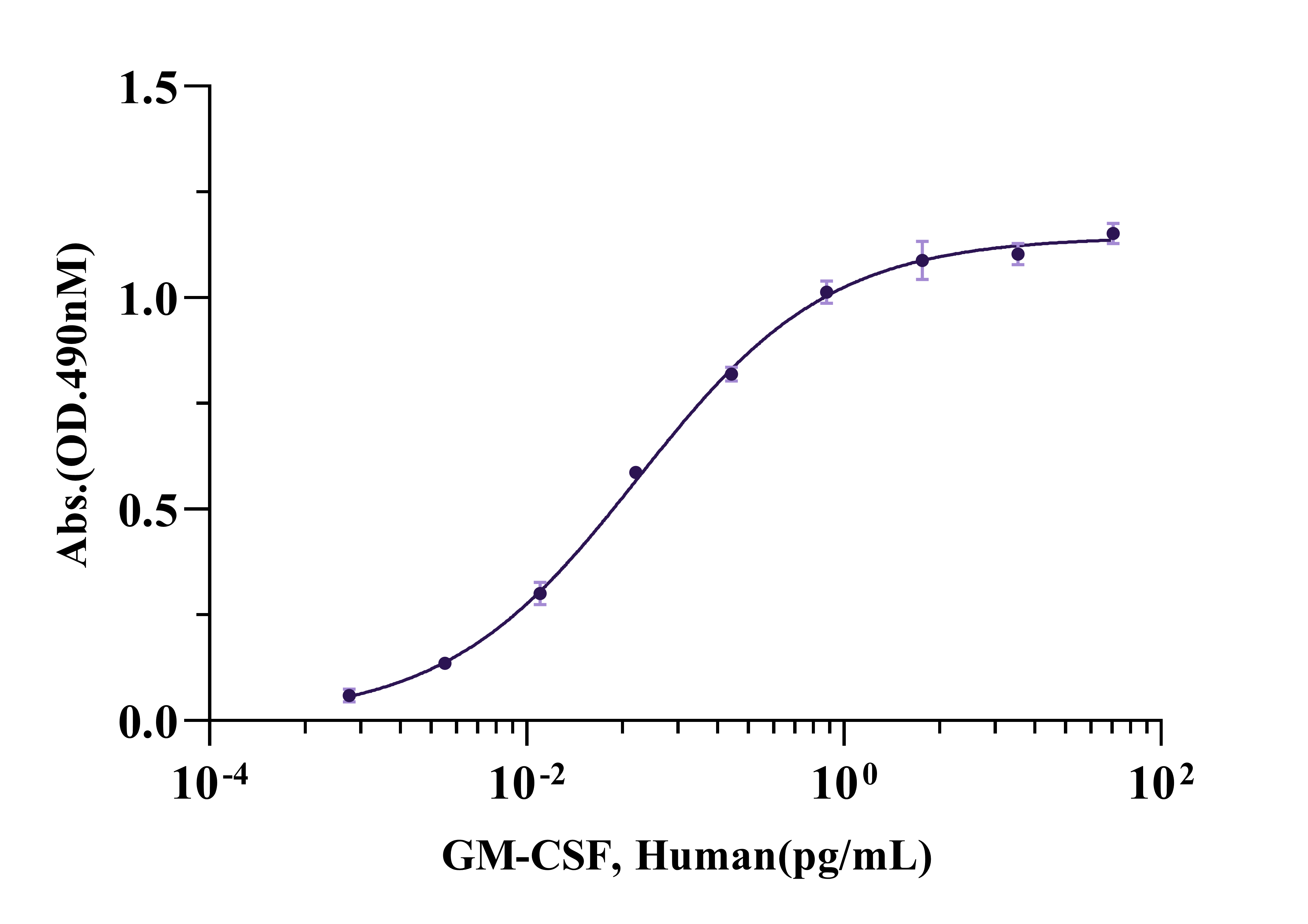
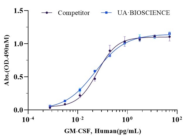
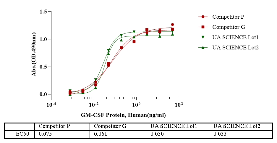
Measured in a cell proliferation assay using TF-1 human erythroleukemic cells. The EC50 for this effect is less than 0.1ng/ml.
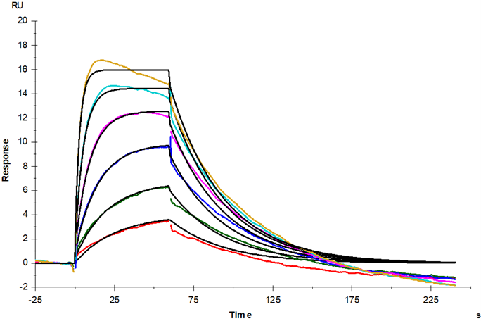
Anti-His antibody Immobilized on CM5 Chip captured GM-CSFR α His Tag, Human (Cat. No. UA010189), can bind GM-CSF, Human (UA040002) with an affinity constant of 11.9 nM as determined in SPR assay.
 您现在的位置:
