
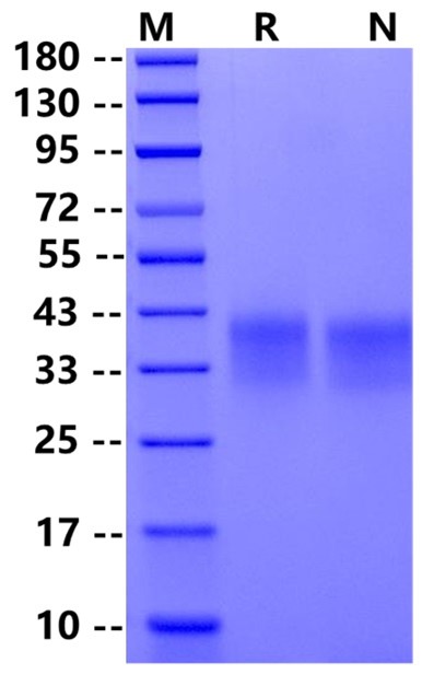
Leu25-Gln167, with C-terminal Avi & 8*His Tag LDSPDRPWNPPTFSPALLVVTEGDNATFTCSFSNTSESFVLNWYRMSPSNQTDKLAAFPEDRSQPGQDCRFRVTQLPNGRDFHMSVVRARRNDSGTYLCGAISLAPKAQIKESLRAELRVTERRAEVPTAHPSPSPRPAGQFQGGGSGLNDIFEAQKIEWHEHHHHHHHH
30-43kDa
Reconstitute at 0.1-1 mg/ml according to the size in ultrapure water after rapid centrifugation.
1、Ishida Y. et al. (1992) Induced expression of PD-1, a novel member of the immunoglobulin gene superfamily, upon programmed cell death. EMBO J. 11: 3887.
Programmed death 1 receptor (PD-1), also known as CD279, is a type I transmembrane protein belonging to the immune regulatory receptor CD28 family. PD-1 is expressed on the surface of activated T cells, B cells, macrophages, myelocytes, and a subset of thymocytes. Mature human PD-1 consists of a 148 amino acid (aa) extracellular region (ECD) with one immunoglobulin-like V-type domain, a 24 aa transmembrane domain, and a 95 aa cytoplasmic region. The tail of the cytoplasmic region contains two tyrosine residues, forming the immune receptor tyrosine inhibitory motif (ITIM) and immune receptor tyrosine switching motif (ITSM), which are important for mediating PD-1 signaling. PD-1 has two ligands, PD-L1 and PD-L2, which are members of the B7 family. PD-L1 is expressed in almost all mouse tumor cell lines, whereas the expression of PD-L2 is more restricted and mainly expressed by DC and a few tumor lines.

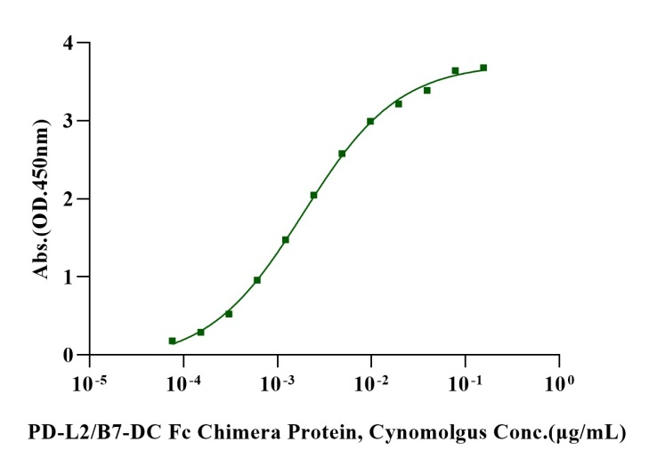
Immobilized Biotinylated PD-1 Avi&His Tag Protein, Human (Cat. No. UA010427) at 2 μg/mL on Streptavidin precoated (0.5μg/well) plate, can bind PD-L2/B7-DC Fc Chimera Protein, Cynomolgus (Cat. No. UA010299) with EC50 of 1.64-2.18ng/ml.
 您现在的位置: