12 months from date of receipt / reconstitution, -20 °C as supplied
| 应用 | 稀释度 |
|---|---|
| FCM | 1:2000 |
| ICC | 1:500 |
CD2 (cluster of differentiation 2) is a cell adhesion molecule found on the surface of T cells and natural killer (NK) cells. It has also been called T-cell surface antigen T11/Leu-5, LFA-2, LFA-3 receptor, erythrocyte receptor and rosette receptor. It interacts with other adhesion molecules, such as lymphocyte function-associated antigen-3 (LFA-3/CD58) in humans, or CD48 in rodents, which are expressed on the surfaces of other cells. CD2 is a specific marker for T cells and NK cells, and can therefore be used in immunohistochemistry to identify the presence of such cells in tissue sections. The great majority of T cell lymphomas and leukaemias also express CD2, making it possible to use the presence of the antigen to distinguish these conditions from B cell neoplasms.
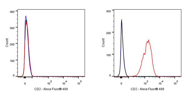
Flow cytometric analysis of Ramos (Human Burkitt's lymphoma B lymphocyte, left) / Jurkat (Human T cell leukemia T lymphocyte, right) cells labelling CD2 antibody at 1/2000 dilution (0.1 μg) / (red) compared with a Mouse monoclonal IgG (Black) isotype control and an unlabelled control (cells without incubation with primary antibody and secondary antibody) (Blue). Goat Anti - Mouse IgG Alexa Fluor® 488 was used as the secondary antibody.
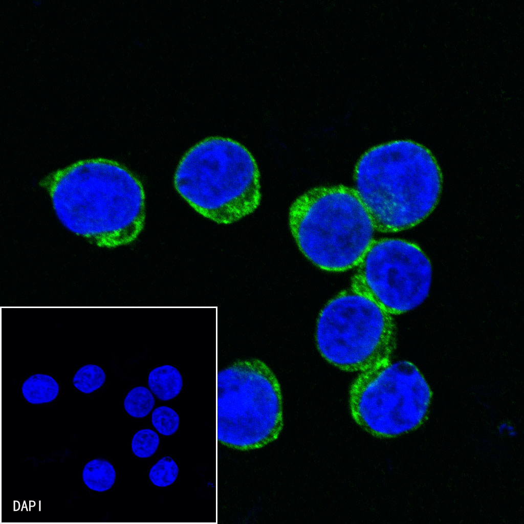
ICC shows positive staining in Jurkat cells. Anti-CD2 antibody was used at 1/500 dilution (Green) and incubated overnight at 4°C. Goat polyclonal Antibody to Mouse IgG - H&L (Alexa Fluor® 488) was used as secondary antibody at 1/1000 dilution. The cells were fixed with 4%PFA and permeabilized with 0.1% PBS-Triton X-100. Nuclei were counterstained with DAPI (Blue).
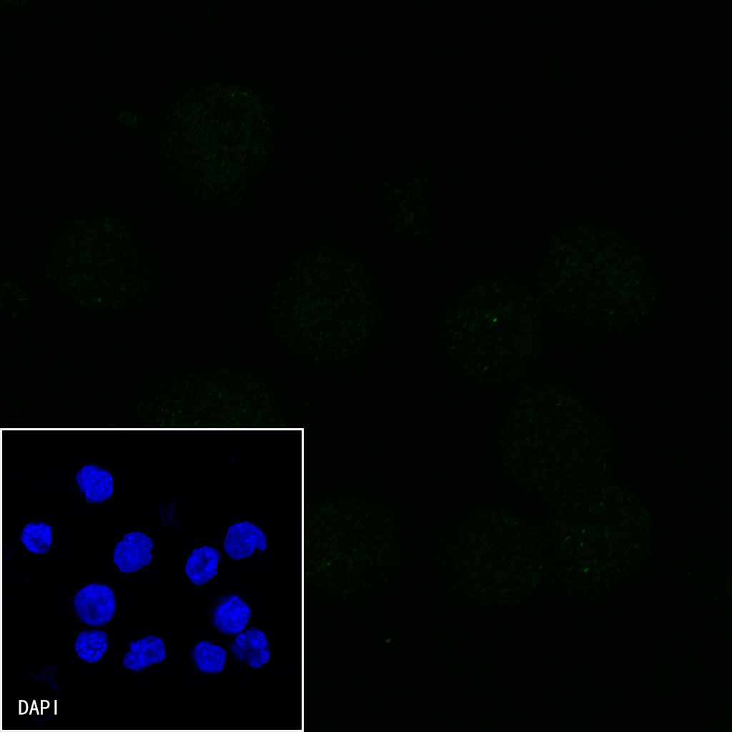
Negative control: ICC shows negative staining in Daudi cells. Anti-CD2 antibody was used at 1/500 dilution and incubated overnight at 4°C. Goat polyclonal Antibody to Mouse IgG - H&L (Alexa Fluor® 488) was used as secondary antibody at 1/1000 dilution. The cells were fixed with 4%PFA and permeabilized with 0.1% PBS-Triton X-100. Nuclei were counterstained with DAPI (Blue).
 您现在的位置: