
12 months from date of receipt / reconstitution, -20 °C as supplied
| 应用 | 稀释度 |
|---|---|
| WB | 1:500 |
| IHC-P | 1:200-1:500 |
| ICC | 1:50 |
| ICFCM | 1:500 |
Forkhead box O3, also known as FOXO3 or FOXO3a, is a human protein encoded by the FOXO3 gene. FOXO3 belongs to the O subclass of the forkhead family of transcription factors which are characterized by a distinct fork head DNA-binding domain. There are three other FoxO family members in humans, FOXO1, FOXO4 and FOXO6. These transcription factors share the ability to be inhibited and translocated out of the nucleus on phosphorylation by proteins such as Akt/PKB in the PI3K signaling pathway (aside from FOXO6, which may be constitutively nuclear). Other post-translational modifications including acetylation and methylation are seen and can result in increased or altered FOXO3a activity.
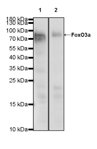
WB result of FoxO3a Rabbit mAb
Primary antibody: FoxO3a Rabbit mAb at 1/500 dilution
Lane 1: HEK-293 whole cell lysate 20 µg
Lane 2: SH-SY5Y whole cell lysate 20 µg
Secondary antibody: Goat Anti-Rabbit IgG, (H+L), HRP conjugated at 1/10000 dilution
Predicted MW: 71 kDa
Observed MW: 82 kDa
(This blot was developed with high sensitivity substrate)
Flow cytometric analysis of 4% PFA fixed 90% methanol permeabilized SH-SY5Y (Human neuroblastoma epithelial cell) cells labelling FoxO3a antibody at 1/500 (0.1 μg) dilution / (red) compared with a Rabbit monoclonal IgG (Black) isotype control and an unlabelled control (cells without incubation with primary antibody and secondary antibody) (Blue). Goat Anti - Rabbit IgG Alexa Fluor® 488 was used as the secondary antibody.
IHC shows positive staining in paraffin-embedded human tonsil. Anti-FoxO3a antibody was used at 1/200 dilution, followed by a HRP Polymer for Mouse & Rabbit IgG (ready to use). Counterstained with hematoxylin. Heat mediated antigen retrieval with Tris/EDTA buffer pH9.0 was performed before commencing with IHC staining protocol.
IHC shows positive staining in paraffin-embedded human testis. Anti-FoxO3a antibody was used at 1/500 dilution, followed by a HRP Polymer for Mouse & Rabbit IgG (ready to use). Counterstained with hematoxylin. Heat mediated antigen retrieval with Tris/EDTA buffer pH9.0 was performed before commencing with IHC staining protocol.
IHC shows positive staining in paraffin-embedded human breast cancer. Anti-FoxO3a antibody was used at 1/500 dilution, followed by a HRP Polymer for Mouse & Rabbit IgG (ready to use). Counterstained with hematoxylin. Heat mediated antigen retrieval with Tris/EDTA buffer pH9.0 was performed before commencing with IHC staining protocol.
IHC shows positive staining in paraffin-embedded human endometrial cancer. Anti-FoxO3a antibody was used at 1/500 dilution, followed by a HRP Polymer for Mouse & Rabbit IgG (ready to use). Counterstained with hematoxylin. Heat mediated antigen retrieval with Tris/EDTA buffer pH9.0 was performed before commencing with IHC staining protocol.
IHC shows positive staining in paraffin-embedded human lung squamous cell carcinoma. Anti-FoxO3a antibody was used at 1/500 dilution, followed by a HRP Polymer for Mouse & Rabbit IgG (ready to use). Counterstained with hematoxylin. Heat mediated antigen retrieval with Tris/EDTA buffer pH9.0 was performed before commencing with IHC staining protocol.
IHC shows positive staining in paraffin-embedded mouse spleen. Anti-FoxO3a antibody was used at 1/500 dilution, followed by a HRP Polymer for Mouse & Rabbit IgG (ready to use). Counterstained with hematoxylin. Heat mediated antigen retrieval with Tris/EDTA buffer pH9.0 was performed before commencing with IHC staining protocol.
IHC shows positive staining in paraffin-embedded rat spleen. Anti-FoxO3a antibody was used at 1/500 dilution, followed by a HRP Polymer for Mouse & Rabbit IgG (ready to use). Counterstained with hematoxylin. Heat mediated antigen retrieval with Tris/EDTA buffer pH9.0 was performed before commencing with IHC staining protocol.
IHC shows positive staining in paraffin-embedded rat stomach. Anti-FoxO3a antibody was used at 1/500 dilution, followed by a HRP Polymer for Mouse & Rabbit IgG (ready to use). Counterstained with hematoxylin. Heat mediated antigen retrieval with Tris/EDTA buffer pH9.0 was performed before commencing with IHC staining protocol.
ICC shows positive staining in SH-SY5Y cells. Anti-FoxO3a antibody was used at 1/50 dilution (Green) and incubated overnight at 4°C. Goat polyclonal Antibody to Rabbit IgG - H&L (Alexa Fluor® 488) was used as secondary antibody at 1/1000 dilution. The cells were fixed with 4% PFA and permeabilized with 0.1% PBS-Triton X-100. Nuclei were counterstained with DAPI (Blue). Counterstain with tubulin (red).
 您现在的位置: