

PBS, 40% Glycerol, 0.05% BSA, 0.03% Proclin 300
12 months from date of receipt / reconstitution, -20 °C as supplied
Isotype control antibodies, to estimate the nonspecific binding of target. Use at concentrations comparable to those of the specific antibody of interest.
WB result of Rabbit mAb IgG Isotype Control
Primary antibody: Rabbit mAb IgG Isotype Control at 1/1000 dilution
Lane 1: HeLa whole cell lysate 20 µg
Lane 2: THP-1 whole cell lysate 20 µg
Secondary antibody: Goat Anti-Rabbit IgG, (H+L), HRP conjugated at 1/10000 dilution
WB result of Rabbit mAb IgG Isotype Control
Primary antibody: Rabbit mAb IgG Isotype Control at 1/1000 dilution
Lane 1: NIH/3T3 whole cell lysate 20 µg
Lane 2: mouse spleen lysate 20 µg
Secondary antibody: Goat Anti-Rabbit IgG, (H+L), HRP conjugated at 1/10000 dilution
WB result of Rabbit mAb IgG Isotype Control
Primary antibody: Rabbit mAb IgG Isotype Control at 1/1000 dilution
Lane 1: C6 whole cell lysate 20 µg
Lane 2: rat spleen lysate 20 µg
Secondary antibody: Goat Anti-Rabbit IgG, (H+L), HRP conjugated at 1/10000 dilution
Flow cytometric analysis of mouse primary splenocytes labeling Rabbit IgG isotype control at 1/50 dilution (1 μg) / (left panel) compared with CD8α antibody at 1/50 (1 μg) dilution (S0B0034) / (right panel). Goat Anti-Rabbit IgG Alexa Fluor 488 was used as the secondary antibody.
Then cells were stained with CD4 - Alexa Fluor 647 separately. CD4 and CD8α are mutually exclusive expressed in mouse primary splenocytes. Gated on total viable cells.
Flow cytometric analysis of 4% PFA fixed 90% methanol permeabilized HeLa (human cervical adenocarcinoma epithelial cell) cells labelling Rabbit IgG isotype control at 1/50 dilution (1 μg) / (Red) compared with a Rabbit monoclonal IgG (Black) isotype control and an unlabelled control (cells without incubation with primary antibody and secondary antibody) (Blue). A Goat Anti-Rabbit IgG Alexa Fluor 488 was used as the secondary antibody.
Flow cytometric analysis of HeLa cells labelling Stat6 antibody at 1/50 (1 μg) dilution/ (red) (S0B0085) compared with a Rabbit monoclonal IgG (Black) isotype control and an unlabelled control (cells without incubation with primary antibody and secondary antibody) (Blue). Goat Anti-Rabbit IgG Alexa Fluor® 488 was used as the secondary antibody.
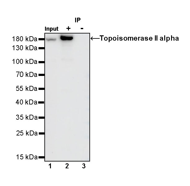
Topoisomerase II alpha Rabbit mAb at 1/50 dilution (1 µg) immunoprecipitating Topoisomerase II alpha in 0.4 mg SK-BR-3 whole cell lysate.
Western blot was performed on the immunoprecipitate using Topoisomerase II alpha Rabbit mAb at 1/1000 dilution.
Secondary antibody (HRP) for IP was used at 1/400 dilution.
Lane 1: SK-BR-3 whole cell lysate 20 µg (Input)
Lane 2: Topoisomerase II alpha Rabbit mAb IP in SK-BR-3 whole cell lysate
Lane 3: Rabbit mAb IgG Isotype Control IP in SK-BR-3 whole cell lysate
Predicted MW: 174 kDa
Observed MW: 174 kDa
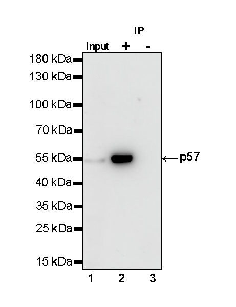
p57 Rabbit mAb at 1/50 dilution (1 µg) immunoprecipitating p57 in 0.4 mg HeLa treated with Dexamethasone (50nM, 16hr) whole cell lysate.
Western blot was performed on the immunoprecipitate using p57 Rabbit mAb at 1/1000 dilution.
Secondary antibody (HRP) for IP was used at 1/400 dilution.
Lane 1: HeLa treated with Dexamethasone (50nM, 16hr) whole cell lysate 10 µg (Input)
Lane 2: p57 Rabbit mAb IP in HeLa treated with Dexamethasone (50nM, 16hr) whole cell lysate
Lane 3: Rabbit mAb IgG Isotype Control IP in HeLa treated with Dexamethasone (50nM, 16hr) whole cell lysate
Predicted MW: 57 kDa
Observed MW: 52 kDa
(This blot was developed with high sensitivity substrate)
IHC shows negative staining in paraffin-embedded mouse cerebral cortex. Rabbit mAb IgG Isotype Control was used at 1/2000 dilution, followed by a HRP Polymer for Mouse & Rabbit IgG (ready to use). Counterstained with hematoxylin. Heat mediated antigen retrieval with Tris/EDTA buffer pH9.0 was performed before commencing with IHC staining protocol.
IHC shows negative staining in paraffin-embedded rat cerebral cortex. Rabbit mAb IgG Isotype Control was used at 1/2000 dilution, followed by a HRP Polymer for Mouse & Rabbit IgG (ready to use). Counterstained with hematoxylin. Heat mediated antigen retrieval with Tris/EDTA buffer pH9.0 was performed before commencing with IHC staining protocol.
IHC shows negative staining in paraffin-embedded human tonsil. Rabbit mAb IgG Isotype Control was used at 1/2000 dilution, followed by a HRP Polymer for Mouse & Rabbit IgG (ready to use). Counterstained with hematoxylin. Heat mediated antigen retrieval with Tris/EDTA buffer pH9.0 was performed before commencing with IHC staining protocol.
IHC shows negative staining in paraffin-embedded human breast cancer. Rabbit mAb IgG Isotype Control was used at 1/2000 dilution, followed by a HRP Polymer for Mouse & Rabbit IgG (ready to use). Counterstained with hematoxylin. Heat mediated antigen retrieval with Tris/EDTA buffer pH9.0 was performed before commencing with IHC staining protocol.
ICC shows negative staining in HeLa cells. Rabbit mAb IgG Isotype Control was used at 1/50 dilution and incubated overnight at 4°C. Goat polyclonal Antibody to Rabbit IgG - H&L (Alexa Fluor® 488) was used as secondary antibody at 1/1000 dilution. The cells were fixed with 4% PFA and permeabilized with 0.1% PBS-Triton X-100. Nuclei were counterstained with DAPI.
ICC shows negative staining in C2C12 cells. Rabbit mAb IgG Isotype Control was used at 1/50 dilution and incubated overnight at 4°C. Goat polyclonal Antibody to Rabbit IgG - H&L (Alexa Fluor® 488) was used as secondary antibody at 1/1000 dilution. The cells were fixed with 4% PFA and permeabilized with 0.1% PBS-Triton X-100. Nuclei were counterstained with DAPI.
ICC shows negative staining in C6 cells. Rabbit mAb IgG Isotype Control was used at 1/50 dilution and incubated overnight at 4°C. Goat polyclonal Antibody to Rabbit IgG - H&L (Alexa Fluor® 488) was used as secondary antibody at 1/1000 dilution. The cells were fixed with 100% ice-cold methanol and permeabilized with 0.1% PBS-Triton X-100. Nuclei were counterstained with DAPI.
 您现在的位置: