
12 months from date of receipt / reconstitution, -20 °C as supplied
| 应用 | 稀释度 |
|---|---|
| WB | 1:1000 |
| IHC | 1:2000 |
| ICFCM | 1:500 |
| ICC | 1:500 |
| IP | 1:50 |
DNA topoisomerase IIα is a human enzyme encoded by the TOP2A gene. Topoisomerase IIα relieves topological DNA stress during transcription, condenses chromosomes, and separates chromatids. It catalyzes the transient breaking and rejoining of two strands of duplex DNA which allows the strands to pass through one another. The gene encoding this enzyme functions as the target for several chemotherapy agents and a variety of mutations in this gene have been associated with the development of drug resistance. Reduced activity of this enzyme may also play a role in ataxia-telangiectasia.
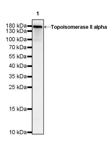
WB result of Topoisomerase II alpha Rabbit mAb
Primary antibody: Topoisomerase II alpha Rabbit mAb at 1/1000 dilution
Lane 1: SK-BR-3 whole cell lysate 20 µg
Secondary antibody: Goat Anti-Rabbit IgG, (H+L), HRP conjugated at 1/10000 dilution
Predicted MW: 174 kDa
Observed MW: 174 kDa
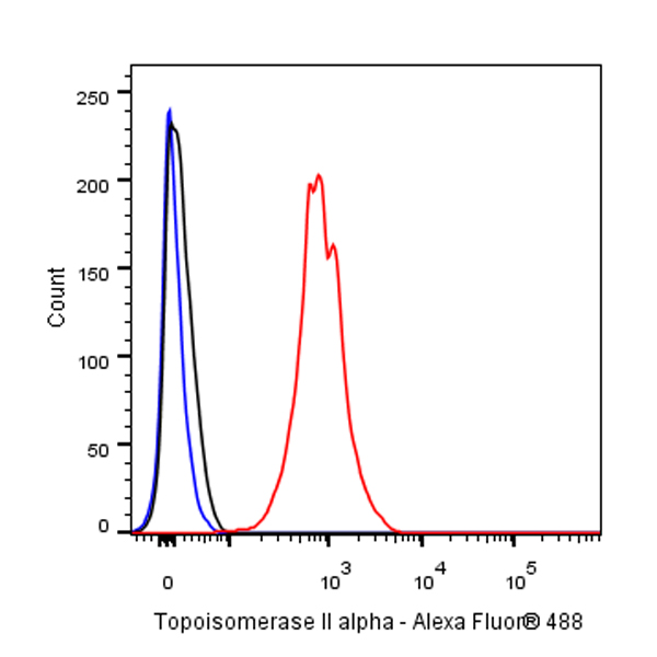
Flow cytometric analysis of 4% PFA fixed 90% methanol permeabilized HeLa (Human cervix adenocarcinoma epithelial cell) cells labeling Topoisomerase II alpha at 1/500 dilution (0.1 μg) / (red) compared with a rabbit monoclonal IgG isotype control (black) and an unlabeled control (cells without incubation with primary antibody and secondary antibody) (Blue). Goat Anti - Rabbit IgG Alexa Fluor® 488 was used as the secondary antibody.

Topoisomerase II alpha Rabbit mAb at 1/50 dilution (1 µg) immunoprecipitating Topoisomerase II alpha in 0.4 mg SK-BR-3 whole cell lysate.
Western blot was performed on the immunoprecipitate using Topoisomerase II alpha Rabbit mAb at 1/1000 dilution.
Secondary antibody (HRP) for IP was used at 1/400 dilution.
Lane 1: SK-BR-3 whole cell lysate 20 µg (Input)
Lane 2: Topoisomerase II alpha Rabbit mAb IP in SK-BR-3 whole cell lysate
Lane 3: Rabbit monoclonal IgG IP in SK-BR-3 whole cell lysate
Predicted MW: 174 kDa
Observed MW: 174 kDa
IHC shows positive staining in paraffin-embedded human colon. Anti-Topoisomerase II alpha antibody was used at 1/2000 dilution, followed by a HRP Polymer for Mouse & Rabbit IgG (ready to use). Counterstained with hematoxylin. Heat mediated antigen retrieval with Tris/EDTA buffer pH9.0 was performed before commencing with IHC staining protocol.
IHC shows positive staining in paraffin-embedded human tonsil. Anti-Topoisomerase II alpha antibody was used at 1/2000 dilution, followed by a HRP Polymer for Mouse & Rabbit IgG (ready to use). Counterstained with hematoxylin. Heat mediated antigen retrieval with Tris/EDTA buffer pH9.0 was performed before commencing with IHC staining protocol.
IHC shows positive staining in paraffin-embedded human testis. Anti-Topoisomerase II alpha antibody was used at 1/2000 dilution, followed by a HRP Polymer for Mouse & Rabbit IgG (ready to use). Counterstained with hematoxylin. Heat mediated antigen retrieval with Tris/EDTA buffer pH9.0 was performed before commencing with IHC staining protocol.
IHC shows positive staining in paraffin-embedded human lung squamous cell carcinoma. Anti-Topoisomerase II alpha antibody was used at 1/2000 dilution, followed by a HRP Polymer for Mouse & Rabbit IgG (ready to use). Counterstained with hematoxylin. Heat mediated antigen retrieval with Tris/EDTA buffer pH9.0 was performed before commencing with IHC staining protocol.
IHC shows positive staining in paraffin-embedded human ovarian carcinoma. Anti-Topoisomerase II alpha antibody was used at 1/2000 dilution, followed by a HRP Polymer for Mouse & Rabbit IgG (ready to use). Counterstained with hematoxylin. Heat mediated antigen retrieval with Tris/EDTA buffer pH9.0 was performed before commencing with IHC staining protocol.
IHC shows positive staining in paraffin-embedded human gastric carcinoma. Anti-Topoisomerase II alpha antibody was used at 1/2000 dilution, followed by a HRP Polymer for Mouse & Rabbit IgG (ready to use). Counterstained with hematoxylin. Heat mediated antigen retrieval with Tris/EDTA buffer pH9.0 was performed before commencing with IHC staining protocol.
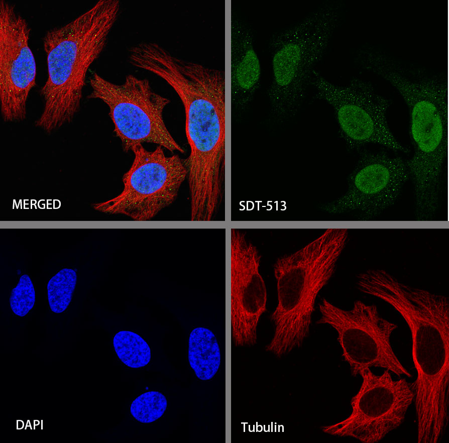
ICC shows positive staining in HeLa cells. Anti-Topoisomerase II alpha antibody was used at 1/500 dilution (Green) and incubated overnight at 4°C. Goat polyclonal Antibody to Rabbit IgG - H&L (Alexa Fluor® 488) was used as secondary antibody at 1/1000 dilution. The cells were fixed with 4%PFA and permeabilized with 0.1% PBS-Triton X-100. Nuclei were counterstained with DAPI (Blue).
 您现在的位置: