
PBS, 40% Glycerol, 0.05%BSA, 0.03% Proclin 300
12 months from date of receipt / reconstitution, -20 °C as supplied
| 应用 | 稀释度 |
|---|---|
| WB | 1:1000 |
| IP | 1:50 |
| IHC | 1:2000 |
| ICC | 1:500 |
| IF | 1:1000 |
| ICFCM | 1:500 |
B7-H3 is a 316 amino acid-long type I transmembrane protein, existing in two isoforms determined by its extracellular domain. In humans it consists of one pair (2Ig-B7-H3) or two identical pairs (4Ig-B7-H3) due to exon duplication. B7-H3 mRNA is expressed in most normal tissues. In contrast, B7-H3 protein has a very limited expression on normal tissues because of its post-transcriptional regulation by microRNAs. However, B7-H3 protein is expressed at high frequency on many different cancer types (60% of all cancers). In non-malignant tissues, B7-H3 has a predominantly inhibitory role in adaptive immunity, suppressing T cell activation and proliferation. In malignant tissues, B7-H3 is an immune checkpoint molecule that inhibits tumor antigen-specific immune responses. B7-H3 also possesses non-immunological pro-tumorigenic functions such as promoting migration, invasion, angiogenesis, chemoresistance, epithelial-to-mesenchymal transition, and affecting tumor cell metabolism.
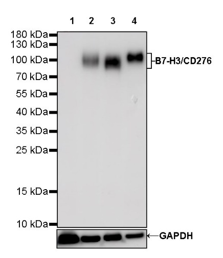
WB result of B7-H3/CD276 Rabbit mAb
Primary antibody: B7-H3/CD276 Rabbit mAb at 1/1000 dilution
Lane 1: Raji whole cell lysate 20 µg
Lane 2: LNCaP whole cell unboiled RIPA lysate 20 µg
Lane 3: HEK-293 whole cell unboiled RIPA lysate 20 µg
Lane 4: U-2 OS whole cell lysate 20 µg
Negative control: Raji whole cell lysate
Secondary antibody: Goat Anti-Rabbit IgG, (H+L), HRP conjugated at 1/10000 dilution
Predicted MW: 90 kDa
Observed MW: 90~110 kDa
Exposure time: 180 s
Flow cytometric analysis of 4% PFA fixed 90% methanol permeabilized LNCaP (Human prostate carcinoma epithelial cell) cells labelling B7-H3/CD276 antibody at 1/500 dilution (0.1 μg) / (red) compared with a Rabbit monoclonal IgG (Black) isotype control and an unlabelled control (cells without incubation with primary antibody and secondary antibody) (Blue). Goat Anti - Rabbit IgG Alexa Fluor® 488 was used as the secondary antibody.
B7-H3/CD276 Rabbit mAb at 1/50 dilution (1 µg) immunoprecipitating B7-H3/CD276 in 0.4 mg LnCap whole cell lysate.
Western blot was performed on the immunoprecipitate using B7-H3/CD276 Rabbit mAb at 1/1000 dilution.
Secondary antibody (HRP) for IP was used at 1/400 dilution.
Lane 1: LnCap whole cell lysate 20 µg (Input)
Lane 2: B7-H3/CD276 Rabbit mAb IP in LnCap whole cell lysate
Lane 3: Rabbit monoclonal IgG IP in LnCap whole cell lysate
Predicted MW: 90 kDa
Observed MW: 90~110 kDa
Exposure time: 60 s
IHC shows positive staining in paraffin-embedded human placenta. Anti- B7-H3/CD276 antibody was used at 1/2000 dilution, followed by a HRP Polymer for Mouse & Rabbit IgG (ready to use). Counterstained with hematoxylin. Heat mediated antigen retrieval with Tris/EDTA buffer pH9.0 was performed before commencing with IHC staining protocol.
IHC shows positive staining in paraffin-embedded human colon. Anti- B7-H3/CD276 antibody was used at 1/2000 dilution, followed by a HRP Polymer for Mouse & Rabbit IgG (ready to use). Counterstained with hematoxylin. Heat mediated antigen retrieval with Tris/EDTA buffer pH9.0 was performed before commencing with IHC staining protocol.
Negative control: IHC shows negative staining in paraffin-embedded human skeletal muscle. Anti- B7-H3/CD276 antibody was used at 1/2000 dilution, followed by a HRP Polymer for Mouse & Rabbit IgG (ready to use). Counterstained with hematoxylin. Heat mediated antigen retrieval with Tris/EDTA buffer pH9.0 was performed before commencing with IHC staining protocol.
IHC shows positive staining in paraffin-embedded human breast cancer. Anti- B7-H3/CD276 antibody was used at 1/2000 dilution, followed by a HRP Polymer for Mouse & Rabbit IgG (ready to use). Counterstained with hematoxylin. Heat mediated antigen retrieval with Tris/EDTA buffer pH9.0 was performed before commencing with IHC staining protocol.
IHC shows positive staining in paraffin-embedded human colon cancer. Anti- B7-H3/CD276 antibody was used at 1/2000 dilution, followed by a HRP Polymer for Mouse & Rabbit IgG (ready to use). Counterstained with hematoxylin. Heat mediated antigen retrieval with Tris/EDTA buffer pH9.0 was performed before commencing with IHC staining protocol.
IHC shows positive staining in paraffin-embedded human endometrial cancer. Anti- B7-H3/CD276 antibody was used at 1/2000 dilution, followed by a HRP Polymer for Mouse & Rabbit IgG (ready to use). Counterstained with hematoxylin. Heat mediated antigen retrieval with Tris/EDTA buffer pH9.0 was performed before commencing with IHC staining protocol.
IHC shows positive staining in paraffin-embedded human lung cancer. Anti- B7-H3/CD276 antibody was used at 1/2000 dilution, followed by a HRP Polymer for Mouse & Rabbit IgG (ready to use). Counterstained with hematoxylin. Heat mediated antigen retrieval with Tris/EDTA buffer pH9.0 was performed before commencing with IHC staining protocol.
IHC shows positive staining in paraffin-embedded human ovarian cancer. Anti- B7-H3/CD276 antibody was used at 1/2000 dilution, followed by a HRP Polymer for Mouse & Rabbit IgG (ready to use). Counterstained with hematoxylin. Heat mediated antigen retrieval with Tris/EDTA buffer pH9.0 was performed before commencing with IHC staining protocol.
ICC shows positive staining in LNCaP cells. Anti- B7-H3/CD276 antibody was used at 1/500 dilution (Green) and incubated overnight at 4°C. Goat polyclonal Antibody to Rabbit IgG - H&L (Alexa Fluor® 488) was used as secondary antibody at 1/1000 dilution. The cells were fixed with 100% ice-cold methanol and permeabilized with 0.1% PBS-Triton X-100. Nuclei were counterstained with DAPI (Blue). Counterstain with tubulin (Red).
IF shows positive staining in paraffin-embedded human lung adenocarcinoma. Anti-B7-H3 antibody was used at 1/1000 dilution (Green) and incubated overnight at 4°C. Goat polyclonal Antibody to Rabbit IgG - H&L (Alexa Fluor® 488) was used as secondary antibody at 1/1000 dilution. Counterstained with DAPI (Blue). Heat mediated antigen retrieval with EDTA buffer pH9.0 was performed before commencing with IF staining protocol.
IF shows positive staining in paraffin-embedded human squamous cell carcinoma. Anti-B7-H3 antibody was used at 1/1000 dilution (Green) and incubated overnight at 4°C. Goat polyclonal Antibody to Rabbit IgG - H&L (Alexa Fluor® 488) was used as secondary antibody at 1/1000 dilution. Counterstained with DAPI (Blue). Heat mediated antigen retrieval with EDTA buffer pH9.0 was performed before commencing with IF staining protocol.
 您现在的位置: