
PBS, 40% Glycerol, 0.05%BSA, 0.03% Proclin 300
12 months from date of receipt / reconstitution, -20 °C as supplied
| 应用 | 稀释度 |
|---|---|
| WB | 1:1000 |
| IP | 1:50 |
| IHC | 1:500-1:2000 |
| ICFCM | 1:500 |
Indoleamine-pyrrole 2,3-dioxygenase (IDO or INDO) is a heme-containing enzyme physiologically expressed in a number of tissues and cells, such as the small intestine, lungs, female genital tract or placenta. IDO is an important part of the immune system and plays a part in natural defense against various pathogens.It is produced by the cells in response to inflammation and has an immunosuppressive function because of its ability to limit T-cell function and engage mechanisms of immune tolerance.Emerging evidence suggests that IDO becomes activated during tumor development, helping malignant cells escape eradication by the immune system. Expression of IDO has been described in a number of types of cancer, such as acute myeloid leukemia, ovarian cancer or colorectal cancer.
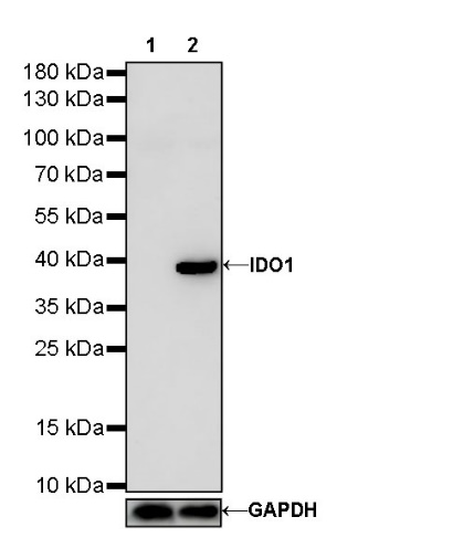
WB result of IDO1/Indoleamine 2,3-dioxygenase Rabbit mAb
Primary antibody: IDO1/Indoleamine 2,3-dioxygenase Rabbit mAb at 1/1000 dilution
Lane 1: HeLa whole cell lysate 20 µg
Lane 2: HeLa treated with IFN-γ (100 ng/mL, 16 hr) whole cell lysate 20 µg
Secondary antibody: Goat Anti-Rabbit IgG, (H+L), HRP conjugated at 1/10000 dilution
Predicted MW: 45 kDa
Observed MW: 40 kDa
WB result of IDO1/Indoleamine 2,3-dioxygenase Rabbit mAb
Primary antibody: IDO1/Indoleamine 2,3-dioxygenase Rabbit mAb at 1/1000 dilution
Lane 1: SK-OV-3 whole cell lysate 20 µg
Secondary antibody: Goat Anti-Rabbit IgG, (H+L), HRP conjugated at 1/10000 dilution
Predicted MW: 45 kDa
Observed MW: 40 kDa
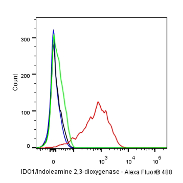
Intracellular flow cytometric analysis of 4% PFA fixed 90% methanol permeabilized HeLa (Mouse myoblasts myoblast), treated with 50ng/ml IFN-γ for 16h (Red) or untreated (Green), labeling IDO1/Indoleamine 2,3-dioxygenase at 1/500 dilution (0.1 μg) compared with a Rabbit monoclonal IgG isotype control (Black) and an unlabeled control (cells without incubation with primary antibody and secondary antibody) (Blue). Goat Anti - Rabbit IgG Alexa Fluor® 488 was used as the secondary antibody.
IDO1/Indoleamine 2,3-dioxygenase Rabbit mAb at 1/50 dilution (1 µg) immunoprecipitating IDO1/Indoleamine 2,3-dioxygenase in 0.4 mg SK-OV-3 whole cell lysate.
Western blot was performed on the immunoprecipitate using IDO1/Indoleamine 2,3-dioxygenase Rabbit mAb at 1/1000 dilution.
Secondary antibody (HRP) for IP was used at 1/400 dilution.
Lane 1: SK-OV-3 whole cell lysate 20 µg (Input)
Lane 2: IDO1/Indoleamine 2,3-dioxygenase Rabbit mAb IP in SK-OV-3 whole cell lysate
Lane 3: Rabbit monoclonal IgG IP in SK-OV-3 whole cell lysate
Predicted MW: 45 kDa
Observed MW: 40 kDa
IHC shows positive staining in paraffin-embedded human placenta. Anti-IDO1/Indoleamine 2,3-dioxygenase antibody was used at 1/500 dilution, followed by a HRP Polymer for Mouse & Rabbit IgG (ready to use). Counterstained with hematoxylin. Heat mediated antigen retrieval with Tris/EDTA buffer pH9.0 was performed before commencing with IHC staining protocol.
IHC shows positive staining in paraffin-embedded human tonsil. Anti-IDO1/Indoleamine 2,3-dioxygenase antibody was used at 1/2000 dilution, followed by a HRP Polymer for Mouse & Rabbit IgG (ready to use). Counterstained with hematoxylin. Heat mediated antigen retrieval with Tris/EDTA buffer pH9.0 was performed before commencing with IHC staining protocol.
IHC shows positive staining in paraffin-embedded human stomach. Anti-IDO1/Indoleamine 2,3-dioxygenase antibody was used at 1/2000 dilution, followed by a HRP Polymer for Mouse & Rabbit IgG (ready to use). Counterstained with hematoxylin. Heat mediated antigen retrieval with Tris/EDTA buffer pH9.0 was performed before commencing with IHC staining protocol.
IHC shows positive staining in paraffin-embedded human endometrial cancer. Anti-IDO1/Indoleamine 2,3-dioxygenase antibody was used at 1/2000 dilution, followed by a HRP Polymer for Mouse & Rabbit IgG (ready to use). Counterstained with hematoxylin. Heat mediated antigen retrieval with Tris/EDTA buffer pH9.0 was performed before commencing with IHC staining protocol.
IHC shows positive staining in paraffin-embedded human Hodgkin’s lymphoma. Anti-IDO1/Indoleamine 2,3-dioxygenase antibody was used at 1/2000 dilution, followed by a HRP Polymer for Mouse & Rabbit IgG (ready to use). Counterstained with hematoxylin. Heat mediated antigen retrieval with Tris/EDTA buffer pH9.0 was performed before commencing with IHC staining protocol.
IHC shows positive staining in paraffin-embedded human diffuse large B-cell lymphoma. Anti-IDO1/Indoleamine 2,3-dioxygenase antibody was used at 1/2000 dilution, followed by a HRP Polymer for Mouse & Rabbit IgG (ready to use). Counterstained with hematoxylin. Heat mediated antigen retrieval with Tris/EDTA buffer pH9.0 was performed before commencing with IHC staining protocol.
 您现在的位置: