
PBS, 40% Glycerol, 0.05%BSA, 0.03% Proclin 300
12 months from date of receipt / reconstitution, -20 °C as supplied
| 应用 | 稀释度 |
|---|---|
| Dot Blot | 1:1000 |
| WB | 1:1000 |
| IP | 1:50 |
| IHC | 1:500 |
| ICC | 1:500 |
| ChIP | 1:20~1:50 |
H3K36me is an epigenetic modification to the DNA packaging protein Histone H3, specifically, the mono-methylation at the 36th lysine residue of the histone H3 protein. The methylation of H3K36 has particularly had effects in transcriptional repression, alternative splicing, dosage compensation, DNA replication and repair, DNA methylation, and the transmission of the memory of gene expression from parents to offspring during development.
WB result of Histone H3 (mono methyl K36) Rabbit mAb
Primary antibody: Histone H3 (mono methyl K36) Rabbit mAb at 1/1000 dilution
Lane 1: HeLa whole cell lysate 20 µg
Lane 2: Jurkat whole cell lysate 20 µg
Secondary antibody: Goat Anti-Rabbit IgG, (H+L), HRP conjugated at 1/10000 dilution
Predicted MW: 17 kDa
Observed MW: 17 kDa
Exposure time: 180 s
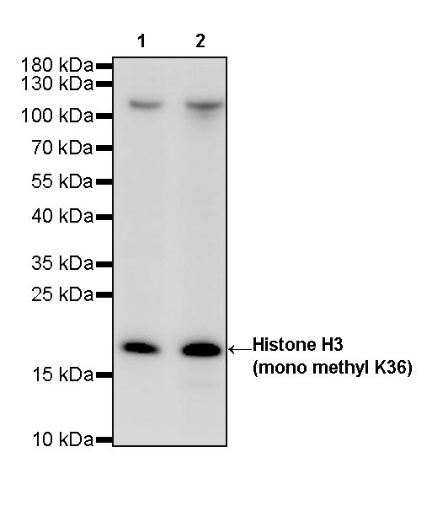
WB result of Histone H3 (mono methyl K36) Rabbit mAb
Primary antibody: Histone H3 (mono methyl K36) Rabbit mAb at 1/1000 dilution
Lane 1: mouse liver lysate 20 µg
Lane 2: mouse kidney lysate 20 µg
Secondary antibody: Goat Anti-Rabbit IgG, (H+L), HRP conjugated at 1/10000 dilution
Predicted MW: 17 kDa
Observed MW: 17 kDa
Exposure time: 180 s
WB result of Histone H3 (mono methyl K36) Rabbit mAb
Primary antibody: Histone H3 (mono methyl K36) Rabbit mAb at 1/1000 dilution
Lane 1: rat liver lysate 20 µg
Secondary antibody: Goat Anti-Rabbit IgG, (H+L), HRP conjugated at 1/10000 dilution
Predicted MW: 17 kDa
Observed MW: 17 kDa
Exposure time: 180 s
Flow cytometric analysis of 4% PFA fixed 90% methanol permeabilized HeLa (Human cervix adenocarcinoma epithelial cell) cells labelling Histone H3 (mono methyl K36) antibody at 1/500 dilution (0.1 μg) / (red) compared with a Rabbit monoclonal IgG (Black) isotype control and an unlabelled control (cells without incubation with primary antibody and secondary antibody) (Blue). Goat Anti - Rabbit IgG Alexa Fluor ® 488 was used as the secondary antibody.
Histone H3 (mono methyl K36) Rabbit mAb at 1/50 dilution (1 µg) immunoprecipitating Histone H3 (mono methyl K36) in 0.4 mg HeLa whole cell lysate.
Western blot was performed on the immunoprecipitate using Histone H3 (mono methyl K36) Rabbit mAb at 1/1000 dilution.
Secondary antibody (HRP) for IP was used at 1/400 dilution.
Lane 1: HeLa whole cell lysate 20 µg (Input)
Lane 2: Histone H3 (mono methyl K36) Rabbit mAb IP in HeLa whole cell lysate
Lane 3: Rabbit monoclonal IgG IP in HeLa whole cell lysate
Predicted MW: 17 kDa
Observed MW: 17 kDa
Exposure time: 80 s
Dot blot result of Histone H3 (mono methyl K36) Rabbit mAb
Lane 1: H3K36me peptide
Lane 2: H3K36me2 peptide
Lane 3: H3K36me3 peptide
Lane 4: H3K36un peptide
Primary antibody: Histone H3 (mono methyl K36) Rabbit mAb at 1/1000 dilution
Secondary antibody: Goat Anti-Rabbit IgG, (H+L), HRP conjugated at 1/10000 dilution
Exposure time: 17 s
IHC shows positive staining in paraffin-embedded human colon. Anti-Histone H3 (mono methyl K36) antibody was used at 1/500 dilution, followed by a HRP Polymer for Mouse & Rabbit IgG (ready to use). Counterstained with hematoxylin. Heat mediated antigen retrieval with Tris/EDTA buffer pH9.0 was performed before commencing with IHC staining protocol.
IHC shows positive staining in paraffin-embedded human kidney. Anti-Histone H3 (mono methyl K36) antibody was used at 1/500 dilution, followed by a HRP Polymer for Mouse & Rabbit IgG (ready to use). Counterstained with hematoxylin. Heat mediated antigen retrieval with Tris/EDTA buffer pH9.0 was performed before commencing with IHC staining protocol.
IHC shows positive staining in paraffin-embedded human breast cancer. Anti-Histone H3 (mono methyl K36) antibody was used at 1/500 dilution, followed by a HRP Polymer for Mouse & Rabbit IgG (ready to use). Counterstained with hematoxylin. Heat mediated antigen retrieval with Tris/EDTA buffer pH9.0 was performed before commencing with IHC staining protocol.
IHC shows positive staining in paraffin-embedded human colon cancer. Anti-Histone H3 (mono methyl K36) antibody was used at 1/500 dilution, followed by a HRP Polymer for Mouse & Rabbit IgG (ready to use). Counterstained with hematoxylin. Heat mediated antigen retrieval with Tris/EDTA buffer pH9.0 was performed before commencing with IHC staining protocol.
IHC shows positive staining in paraffin-embedded human lung squamous cell carcinoma. Anti-Histone H3 (mono methyl K36) antibody was used at 1/500 dilution, followed by a HRP Polymer for Mouse & Rabbit IgG (ready to use). Counterstained with hematoxylin. Heat mediated antigen retrieval with Tris/EDTA buffer pH9.0 was performed before commencing with IHC staining protocol.
IHC shows positive staining in paraffin-embedded mouse liver. Anti-Histone H3 (mono methyl K36) antibody was used at 1/500 dilution, followed by a HRP Polymer for Mouse & Rabbit IgG (ready to use). Counterstained with hematoxylin. Heat mediated antigen retrieval with Tris/EDTA buffer pH9.0 was performed before commencing with IHC staining protocol.
IHC shows positive staining in paraffin-embedded rat stomach. Anti-Histone H3 (mono methyl K36) antibody was used at 1/500 dilution, followed by a HRP Polymer for Mouse & Rabbit IgG (ready to use). Counterstained with hematoxylin. Heat mediated antigen retrieval with Tris/EDTA buffer pH9.0 was performed before commencing with IHC staining protocol.
ICC shows positive staining in HeLa cells. Anti- Histone H3 (mono methyl K36) antibody was used at 1/500 dilution (Green) and incubated overnight at 4°C. Goat polyclonal Antibody to Rabbit IgG - H&L (Alexa Fluor® 488) was used as secondary antibody at 1/1000 dilution. The cells were fixed with 100% ice-cold methanol and permeabilized with 0.1% PBS-Triton X-100. Nuclei were counterstained with DAPI (Blue).
Chromatin immunoprecipitation (ChIP) was performed on HeLa cells cross - linked with 1% formaldehyde for 10 min, then chromatin was fragmented by sonication. Parallel reactions used Histone H3 (mono methyl K36) Recombinant Rabbit mAb (S-R211) and Rabbit mAb IgG Isotype Control (SDT-R173) at 1:50 for immunoprecipitation. Post - immunoprecipitation, both samples were washed, eluted, and cross - links reversed. Purified DNA was analyzed by qPCR.
qPCR (%input: immunoprecipitated DNA/input DNA) showed the enrichment of RPL30, GAPDH, MYOD1, AFM, SAT-α and SAT-2 in Histone H3 (mono methyl K36) Recombinant Rabbit mAb (S-R211) - immunoprecipitated sample.
 您现在的位置: