
12 months from date of receipt / reconstitution, -20 °C as supplied
| 应用 | 稀释度 |
|---|---|
| WB | 1:200 |
| IP | 1:50 |
| IHC | 1:400 |
| ICC | 1:50 |
B7-H4 is a transmembrane protein that binds an unknown receptor on activated T cells resulting in inhibition of T-cell effector function via cell cycle arrest, decreased proliferation, and reduced IL-2 production. B7-H4 is up-regulated on the surface of cancer cells and immunosuppressive tumor-associated macrophages (TAMs) in a variety of human cancers. Notably, B7-H4 expression levels inversely correlate with patient survival in ovarian cancer, cholangiocarcinoma, breast cancer and endometrial cancer, making B7-H4 an attractive candidate for therapeutic intervention.
WB result of B7-H4 Rabbit mAb
Primary antibody: B7-H4 Rabbit mAb at 1/200 dilution
Lane 1: HeLa whole cell lysate 20 µg
Lane 2: SK-BR-3 whole cell lysate 20 µg
Lane 3: T-47D whole cell lysate 20 µg
Negative control: HeLa whole cell lysate
Secondary antibody: Goat Anti-Rabbit IgG, (H+L), HRP conjugated at 1/10000 dilution
Predicted MW: 31 kDa
Observed MW: 65, 45 kDa
(This blot was developed with high sensitivity substrate)
WB result of B7-H4 Rabbit mAb
Primary antibody: B7-H4 Rabbit mAb at 1/200 dilution
Lane 1: recombinant human B7-H1 protein 10ng (CAT.NO.S0A6006)
Lane 2: recombinant human B7-H2 protein 10ng (CAT.NO.UA010058)
Lane 3: recombinant human B7-H3 protein 10ng (CAT.NO.UA010010)
Lane 4: recombinant human B7-H4 protein 10ng (CAT.NO.UA010121)
Secondary antibody: Goat Anti-Rabbit IgG, (H+L), HRP conjugated at 1/10000 dilution
Predicted MW: 31 kDa
Observed MW: 43~55 kDa
(This blot was developed with high sensitivity substrate)
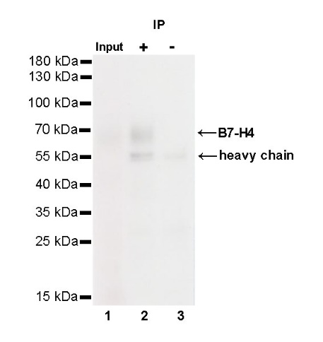
B7-H4 Rabbit mAb at 1/50 dilution (1 µg) immunoprecipitating B7-H4 in 0.4 mg SK-BR-3 whole cell lysate. Western blot was performed on the immunoprecipitate using B7-H4 Rabbit mAb at 1/1000 dilution. Secondary antibody (HRP) for IP was used at 1/400 dilution. Lane 1: SK-BR-3 whole cell lysate 20 µg (Input) Lane 2: B7-H4 Rabbit mAb IP in SK-BR-3 whole cell lysate Lane 3: Rabbit monoclonal IgG IP in SK-BR-3 whole cell lysate Predicted MW: 45 kDa Observed MW: 65 kDa
IHC shows positive staining in paraffin-embedded human breast. Anti-B7-H4 antibody was used at 1/400 dilution, followed by a HRP Polymer for Mouse & Rabbit IgG (ready to use). Counterstained with hematoxylin. Heat mediated antigen retrieval with Tris/EDTA buffer pH9.0 was performed before commencing with IHC staining protocol.
IHC shows positive staining in paraffin-embedded human breast cancer. Anti-B7-H4 antibody was used at 1/400 dilution, followed by a HRP Polymer for Mouse & Rabbit IgG (ready to use). Counterstained with hematoxylin. Heat mediated antigen retrieval with Tris/EDTA buffer pH9.0 was performed before commencing with IHC staining protocol.
IHC shows positive staining in paraffin-embedded human breast cancer. Anti-B7-H4 antibody was used at 1/400 dilution, followed by a HRP Polymer for Mouse & Rabbit IgG (ready to use). Counterstained with hematoxylin. Heat mediated antigen retrieval with Tris/EDTA buffer pH9.0 was performed before commencing with IHC staining protocol.
IHC shows positive staining in paraffin-embedded human breast cancer. Anti-B7-H4 antibody was used at 1/400 dilution, followed by a HRP Polymer for Mouse & Rabbit IgG (ready to use). Counterstained with hematoxylin. Heat mediated antigen retrieval with Tris/EDTA buffer pH9.0 was performed before commencing with IHC staining protocol.
IHC shows positive staining in paraffin-embedded human ovarian cancer. Anti-B7-H4 antibody was used at 1/400 dilution, followed by a HRP Polymer for Mouse & Rabbit IgG (ready to use). Counterstained with hematoxylin. Heat mediated antigen retrieval with Tris/EDTA buffer pH9.0 was performed before commencing with IHC staining protocol.
ICC shows positive staining in SK-BR-3 cells. Anti-B7-H4 antibody was used at 1/50 dilution (Green) and incubated overnight at 4°C. Goat polyclonal Antibody to Rabbit IgG - H&L (Alexa Fluor® 488) was used as secondary antibody at 1/1000 dilution. The cells were fixed with 100% ice-cold methanol and permeabilized with 0.1% PBS-Triton X-100. Nuclei were counterstained with DAPI.
Negative control: ICC shows negative staining in HeLa cells. Anti-B7-H4 antibody was used at 1/50 dilution and incubated overnight at 4°C. Goat polyclonal Antibody to Rabbit IgG - H&L (Alexa Fluor® 488) was used as secondary antibody at 1/1000 dilution. The cells were fixed with 100% ice-cold methanol and permeabilized with 0.1% PBS-Triton X-100. Nuclei were counterstained with DAPI.
 您现在的位置: