
12 months from date of receipt / reconstitution, -20 °C as supplied
| 应用 | 稀释度 |
|---|---|
| WB | 1:1000-1:2000 |
| IHC | 1:2000 |
| ICC | 1:500 |
| ICFCM | 1:500 |
Lumican, also known as LUM, is an extracellular matrix protein that, in humans, is encoded by the LUM gene on chromosome 12 [PMID: 7558030]. Lumican is a major keratan sulfate proteoglycan of the cornea but is ubiquitously distributed in most mesenchymal tissues throughout the body [PMID: 12975607]. Lumican is involved in collagen fibril organization and circumferential growth, corneal transparency, and epithelial cell migration and tissue repair. Corneal transparency is possible due to the exact alignment of collagen fibers by lumican (and keratocan) in the intrafibrillar space.
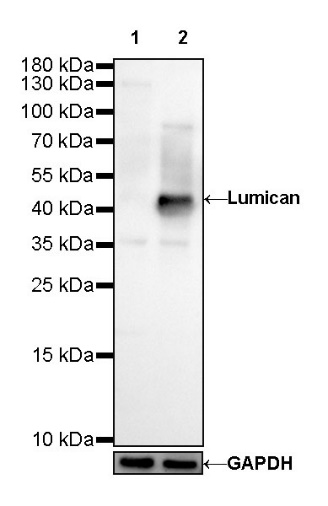
WB result of Lumican Rabbit mAb Primary antibody: Lumican Rabbit mAb at 1/1000 dilution Lane 1: 293T whole cell lysate 20 µg Lane 2: saos-2 whole cell lysate 20 µg Negative control: 293T whole cell lysate Secondary antibody: Goat Anti-Rabbit IgG, (H+L), HRP conjugated at 1/10000 dilution Predicted MW: 38 kDa Observed MW: 50 kDa
WB result of Lumican Rabbit mAb Primary antibody: Lumican Rabbit mAb at 1/2000 dilution Lane 1: mouse skin lysate 20 µg Secondary antibody: Goat Anti-Rabbit IgG, (H+L), HRP conjugated at 1/10000 dilution Predicted MW: 38 kDa Observed MW: 50~70 kDa
WB result of Lumican Rabbit mAb Primary antibody: Lumican Rabbit mAb at 1/2000 dilution Lane 1: rat skin lysate 20 µg Secondary antibody: Goat Anti-Rabbit IgG, (H+L), HRP conjugated at 1/10000 dilution Predicted MW: 38 kDa Observed MW: 60~70 kDa
Flow cytometric analysis of 4% PFA fixed 90% methanol permeabilized 293T (Human embryonic kidney epithelial cell) labelling Lumican antibody at 1/500 (0.1 μg) dilution/ (red) compared with a Rabbit monoclonal IgG (Black) isotype control and an unlabelled control (cells without incubation with primary antibody and secondary antibody) (Blue). Goat Anti-Rabbit IgG Alexa Fluor 488 was used as the secondary antibody.
IHC shows positive staining in paraffin-embedded human colon. Anti-Lumican antibody was used at 1/2000 dilution, followed by a HRP Polymer for Mouse & Rabbit IgG (ready to use). Counterstained with hematoxylin. Heat mediated antigen retrieval with Tris/EDTA buffer pH9.0 was performed before commencing with IHC staining protocol.
IHC shows positive staining in paraffin-embedded human renal clear cell carcinoma. Anti-Lumican antibody was used at 1/2000 dilution, followed by a HRP Polymer for Mouse & Rabbit IgG (ready to use). Counterstained with hematoxylin. Heat mediated antigen retrieval with Tris/EDTA buffer pH9.0 was performed before commencing with IHC staining protocol.
IHC shows positive staining in paraffin-embedded human pancreatic carcinoma. Anti-Lumican antibody was used at 1/2000 dilution, followed by a HRP Polymer for Mouse & Rabbit IgG (ready to use). Counterstained with hematoxylin. Heat mediated antigen retrieval with Tris/EDTA buffer pH9.0 was performed before commencing with IHC staining protocol.
IHC shows positive staining in paraffin-embedded human lung adenocarcinoma. Anti-Lumican antibody was used at 1/2000 dilution, followed by a HRP Polymer for Mouse & Rabbit IgG (ready to use). Counterstained with hematoxylin. Heat mediated antigen retrieval with Tris/EDTA buffer pH9.0 was performed before commencing with IHC staining protocol.
IHC shows positive staining in paraffin-embedded mouse kidney. Anti-Lumican antibody was used at 1/2000 dilution, followed by a HRP Polymer for Mouse & Rabbit IgG (ready to use). Counterstained with hematoxylin. Heat mediated antigen retrieval with Tris/EDTA buffer pH9.0 was performed before commencing with IHC staining protocol.
IHC shows positive staining in paraffin-embedded rat colon. Anti-Lumican antibody was used at 1/2000 dilution, followed by a HRP Polymer for Mouse & Rabbit IgG (ready to use). Counterstained with hematoxylin. Heat mediated antigen retrieval with Tris/EDTA buffer pH9.0 was performed before commencing with IHC staining protocol.
ICC shows positive staining in 293T cells. Anti- Lumican antibody was used at 1/500 dilution (Green) and incubated overnight at 4°C. Goat polyclonal Antibody to Rabbit IgG - H&L (Alexa Fluor® 488) was used as secondary antibody at 1/1000 dilution. The cells were fixed with 4% PFA and permeabilized with 0.1% PBS-Triton X-100. Nuclei were counterstained with DAPI (Blue).
 您现在的位置: