
12 months from date of receipt / reconstitution, -20 °C as supplied
| 应用 | 稀释度 |
|---|---|
| WB | 1:5000 |
| IHC | 1:250 |
| ICC | 1:250 |
| ICFCM | 1:500 |
Ewing’s sarcoma (EWS) is a bone cancer arising predominantly in young children. EWSR1 (Ewing Sarcoma breakpoint region 1/EWS RNA binding protein 1) gene is ubiquitously expressed in most cell types, indicating it has diverse roles in various cellular processes and organ development. Recently, several studies have shown that missense mutations of EWSR1 genes are known to be associated with central nervous system disorders such as amyotrophic lateral sclerosis (ALS) and frontotemporal dementia (FTD). Otherwise, EWSR1 plays epigenetic roles in gene expression, RNA processing, and cellular signal transduction. Interestingly, EWSR1 controls micro RNA (miRNA) levels via Drosha, leading to autophagy dysfunction and impaired dermal development. Ewsr1 deficiency also leads to premature senescence of blood cells and gamete cells with a high rate of apoptosis due to the abnormal meiosis [PMID: 30481590].
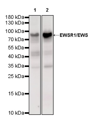
WB result of EWSR1/EWS Rat mAb Primary antibody: EWSR1/EWS Rat mAb at 1/5000 dilution Lane 1: HeLa whole cell lysate 20 µg Lane 2: Jurkat whole cell lysate 20 µg Secondary antibody: Goat Anti-Rat IgG, (H+L), HRP conjugated at 1/10000 dilution Predicted MW: 68 kDa Observed MW: 90 kDa
WB result of EWSR1/EWS Rat mAb Primary antibody: EWSR1/EWS Rat mAb at 1/5000 dilution Lane 1: C6 whole cell lysate 20 µg Secondary antibody: Goat Anti-Rat IgG, (H+L), HRP conjugated at 1/10000 dilution Predicted MW: 68 kDa Observed MW: 90 kDa
Flow cytometric analysis of 4% PFA fixed 90% methanol permeabilized HeLa (Human cervix adenocarcinoma epithelial cell) labelling EWSR1 antibody at 1/500 (0.2 μg) dilution / (red) compared with a Rat monoclonal IgG (Black) isotype control and an unlabelled control (cells without incubation with primary antibody and secondary antibody) (Blue). Goat Anti-Rat IgG Alexa Fluor 488 was used as the secondary antibody.
IHC shows positive staining in paraffin-embedded human colon. Anti-EWSR1/EWS antibody was used at 1/250 dilution, followed by a HRP Polymer for Goat Anti-Rat IgG (H+L). Counterstained with hematoxylin. Heat mediated antigen retrieval with Tris/EDTA buffer pH9.0 was performed before commencing with IHC staining protocol.
IHC shows positive staining in paraffin-embedded human testis. Anti-EWSR1/EWS antibody was used at 1/250 dilution, followed by a HRP Polymer for Goat Anti-Rat IgG (H+L). Counterstained with hematoxylin. Heat mediated antigen retrieval with Tris/EDTA buffer pH9.0 was performed before commencing with IHC staining protocol.
IHC shows positive staining in paraffin-embedded human colon cancer. Anti-EWSR1/EWS antibody was used at 1/250 dilution, followed by a HRP Polymer for Goat Anti-Rat IgG (H+L). Counterstained with hematoxylin. Heat mediated antigen retrieval with Tris/EDTA buffer pH9.0 was performed before commencing with IHC staining protocol.
IHC shows positive staining in paraffin-embedded mouse liver. Anti-EWSR1/EWS antibody was used at 1/250 dilution, followed by a HRP Polymer for Goat Anti-Rat IgG (H+L). Counterstained with hematoxylin. Heat mediated antigen retrieval with Tris/EDTA buffer pH9.0 was performed before commencing with IHC staining protocol.
IHC shows positive staining in paraffin-embedded mouse testis. Anti-EWSR1/EWS antibody was used at 1/250 dilution, followed by a HRP Polymer for Goat Anti-Rat IgG (H+L). Counterstained with hematoxylin. Heat mediated antigen retrieval with Tris/EDTA buffer pH9.0 was performed before commencing with IHC staining protocol.
IHC shows positive staining in paraffin-embedded rat liver. Anti-EWSR1/EWS antibody was used at 1/250 dilution, followed by a HRP Polymer for Goat Anti-Rat IgG (H+L). Counterstained with hematoxylin. Heat mediated antigen retrieval with Tris/EDTA buffer pH9.0 was performed before commencing with IHC staining protocol.
IHC shows positive staining in paraffin-embedded rat testis. Anti-EWSR1/EWS antibody was used at 1/250 dilution, followed by a HRP Polymer for Goat Anti-Rat IgG (H+L). Counterstained with hematoxylin. Heat mediated antigen retrieval with Tris/EDTA buffer pH9.0 was performed before commencing with IHC staining protocol.
ICC shows positive staining in HeLa cells. Anti- EWSR1/EWS antibody was used at 1/250 dilution (Green) and incubated overnight at 4°C. Goat polyclonal Antibody to Rat IgG - H&L (Alexa Fluor® 488) was used as secondary antibody at 1/1000 dilution. The cells were fixed with 4% PFA and permeabilized with 0.1% PBS-Triton X-100. Nuclei were counterstained with DAPI.
 您现在的位置: