
PBS, 40% Glycerol, 0.05% BSA, 0.03% Proclin 300
12 months from date of receipt / reconstitution, -20 °C as supplied
| 应用 | 稀释度 |
|---|---|
| WB | 1:1000 |
| IP | 1:50 |
| IHC | 1:100 |
| ICC | 1:500 |
A single cluster of seven human GBP genes (GBP1-GBP7) is found on chromosome 1q22.2. Human GBP1 is secreted from cells without the need of a leader peptide, and has been shown to exhibit antiviral activity against Vesicular stomatitis virus and Encephalomyocarditis virus, as well as being able to regulate the inhibition of proliferation and invasion of endothelial cells in response to IFN-gamma [PMID: 16936281].
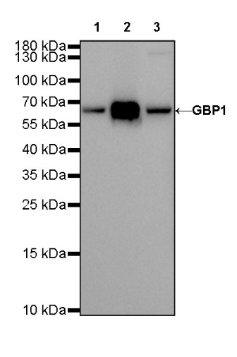
WB result of GBP1 Rat mAb
Primary antibody: GBP1 Rat mAb at 1/1000 dilution
Lane 1: HeLa whole cell lysate 20 µg
Lane 2: HeLa treated with IFN-γ (50 ng/ml 16 hr) whole cell lysate 20 µg
Lane 3: Jurkat whole cell lysate 20 µg
Secondary antibody: Goat Anti-Rat IgG, (H+L), HRP conjugated at 1/10000 dilution
Predicted MW: 68 kDa
Observed MW: 68 kDa
Exposure time: 20 s
GBP1 Rat mAb at 1/200 dilution (1 µg) immunoprecipitating GBP1 in 0.4 mg Jurkat whole cell lysate.
Western blot was performed on the immunoprecipitate using GBP1 Rat mAb at 1/1000 dilution.
Goat Anti-Rat IgG, (H+L), HRP conjugated at 1/10000 dilution.
Lane 1: Jurkat whole cell lysate 20 µg (Input)
Lane 2: GBP1 Rat mAb IP in Jurkat whole cell lysate
Lane 3: Rat monoclonal IgG1 IP in Jurkat whole cell lysate
Predicted MW: 68 kDa
Observed MW: 68 kDa
IHC shows positive staining in paraffin-embedded human tonsil. Anti-GBP1 antibody was used at 1/100 dilution, followed by a Goat Anti-Rat IgG (H+L). Counterstained with hematoxylin. Heat mediated antigen retrieval with Tris/EDTA buffer pH9.0 was performed before commencing with IHC staining protocol.
IHC shows positive staining in paraffin-embedded human spleen. Anti-GBP1 antibody was used at 1/100 dilution, followed by a Goat Anti-Rat IgG (H+L). Counterstained with hematoxylin. Heat mediated antigen retrieval with Tris/EDTA buffer pH9.0 was performed before commencing with IHC staining protocol.
ICC analysis of HeLa cells treated with IFNγ (50ng/ml, 16h) (top panel) and untreated HeLa cells (below panel). Anti- GBP1 antibody was used at 1/500 dilution (Green) and incubated overnight at 4°C. Goat polyclonal Antibody to Rabbit IgG - H&L (Alexa Fluor® 488) was used as secondary antibody at 1/1000 dilution. The cells were fixed with 100% ice-cold methanol and permeabilized with 0.1% PBS-Triton X-100. Nuclei were counterstained with DAPI (Blue).
 您现在的位置: