
| 应用 | 稀释度 |
|---|---|
| WB | 1:1000 |
| IP | 1:50 |
| IHC | 1:500 |
| ICC | 1:500 |
| FCM | 1:500 |
Leukocyte-associated immunoglobulin-like receptor 1 is a protein that in humans is encoded by the LAIR1 gene [PMID: 9285412]. LAIR1 has also been designated as CD305 (cluster of differentiation 305). LAIR-1 is a 32 kDa transmembrane glycoprotein with a single immunoglobulin-like domain and a cytoplasmic tail containing two immune receptor tyrosine-based inhibitory motifs. LAIR-1 recruits SHP-1 and SHP-2 phosphatases upon activation, and cross-linking of the LAIR-1 antigen on natural killer (NK) cells results in strong inhibition of NK cell-mediated cytotoxicity [PMID: 9285412].
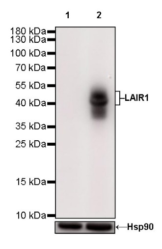
WB result of LAIR1 Rabbit mAb Primary antibody: LAIR1 Rabbit mAb at 1/1000 dilution Lane 1: HeLa whole cell lysate 20 µg Lane 2: THP-1 whole cell lysate 20 µg Negative control: HeLa whole cell lysate Secondary antibody: Goat Anti-Rabbit IgG, (H+L), HRP conjugated at 1/10000 dilution Predicted MW: 31 kDa Observed MW: 40~50 kDa
Flow cytometric analysis of HeLa (Human cervix adenocarcinoma epithelial cell, left) / THP-1 (Human monocytic leukemia monocyte, right) cells labelling LAIR1 antibody at 1/500 dilution (0.1 μg) / (red) compared with a Rabbit monoclonal IgG (Black) isotype control and an unlabelled control (cells without incubation with primary antibody and secondary antibody) (Blue). Goat Anti-Rabbit IgG Alexa Fluor 488 was used as the secondary antibody. Negative control: HeLa
LAIR1 Rabbit mAb at 1/50 dilution (1µg) immunoprecipitating LAIR1 in 0.4 mg THP-1 whole cell lysate. Western blot was performed on the immunoprecipitate using LAIR1 Rabbit mAb at 1/1000 dilution. Secondary antibody (HRP) for IP was used at 1/400 dilution. Lane 1: THP-1 whole cell lysate 20µg (Input) Lane 2: LAIR1 Rabbit mAb IP in THP-1 whole cell lysate Lane 3: Rabbit monoclonal IgG IP in THP-1 whole cell lysate Predicted MW: 40 kDa Observed MW: 40 kDa
IHC shows positive staining in paraffin-embedded human tonsil. Anti-LAIR1 antibody was used at 1/500 dilution, followed by a HRP Polymer for Mouse & Rabbit IgG (ready to use). Counterstained with hematoxylin. Heat mediated antigen retrieval with Tris/EDTA buffer pH9.0 was performed before commencing with IHC staining protocol.
IHC shows positive staining in paraffin-embedded human stomach. Anti-LAIR1 antibody was used at 1/500 dilution, followed by a HRP Polymer for Mouse & Rabbit IgG (ready to use). Counterstained with hematoxylin. Heat mediated antigen retrieval with Tris/EDTA buffer pH9.0 was performed before commencing with IHC staining protocol.
IHC shows positive staining in paraffin-embedded human ovarian cancer. Anti-LAIR1 antibody was used at 1/500 dilution, followed by a HRP Polymer for Mouse & Rabbit IgG (ready to use). Counterstained with hematoxylin. Heat mediated antigen retrieval with Tris/EDTA buffer pH9.0 was performed before commencing with IHC staining protocol.
IHC shows positive staining in paraffin-embedded human gastric cancer. Anti-LAIR1 antibody was used at 1/500 dilution, followed by a HRP Polymer for Mouse & Rabbit IgG (ready to use). Counterstained with hematoxylin. Heat mediated antigen retrieval with Tris/EDTA buffer pH9.0 was performed before commencing with IHC staining protocol.
IHC shows positive staining in paraffin-embedded human diffuse large B-cell lymphoma. Anti-LAIR1 antibody was used at 1/500 dilution, followed by a HRP Polymer for Mouse & Rabbit IgG (ready to use). Counterstained with hematoxylin. Heat mediated antigen retrieval with Tris/EDTA buffer pH9.0 was performed before commencing with IHC staining protocol.
IHC shows positive staining in paraffin-embedded human Hodgkin's lymphoma. Anti-LAIR1 antibody was used at 1/500 dilution, followed by a HRP Polymer for Mouse & Rabbit IgG (ready to use). Counterstained with hematoxylin. Heat mediated antigen retrieval with Tris/EDTA buffer pH9.0 was performed before commencing with IHC staining protocol.
ICC shows positive staining in THP-1 cells. Anti-LAIR1 antibody was used at 1/500 dilution (Green) and incubated overnight at 4°C. Goat polyclonal Antibody to Rabbit IgG - H&L (Alexa Fluor® 488) was used as secondary antibody at 1/1000 dilution. The cells were fixed with 100% ice-cold methanol and permeabilized with 0.1% PBS-Triton X-100. Nuclei were counterstained with DAPI (Blue).
 您现在的位置: