
12 months from date of receipt / reconstitution, -20 °C as supplied
| 应用 | 稀释度 |
|---|---|
| WB | 1:1000 |
| IHC | 1:1000 |
| ICC | 1:500 |
| ICFCM | 1:50 |
E-cadherin is a transmembrane glycoprotein which connects epithelial cells together at adherens junctions. In normal cells, E-cadherin exerts its tumour suppressing role mainly by sequestering β-catenin from its binding to LEF (Lymphoid enhancer factor)/TCF (T cell factor) which serves the function of transcribing genes of the proliferative Wnt signaling pathway. Despite the ongoing debate on whether the loss of E-cadherin is the cause or effect of epithelial-mesenchymal transition (EMT), E-cadherin functional loss has frequently been associated with poor prognosis and survival in patients of various cancers. The dysregulation of E-cadherin expression that leads to carcinogenesis happens mostly at the epigenetic level but there are cases of genetic alterations as well. E-cadherin expression has been linked to the cellular functions of invasiveness reduction, growth inhibition, apoptosis, cell cycle arrest and differentiation.
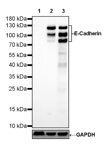
WB result of E-Cadherin Rabbit mAb Primary antibody: E-Cadherin Rabbit mAb at 1/1000 dilution Lane 1: MDA-MB-231 whole cell lysate 20 µg Lane 2: MCF7 whole cell lysate 20 µg Lane 3: HT-29 whole cell lysate 20 µg Negative control: MDA-MB-231 whole cell lysate Secondary antibody: Goat Anti-Rabbit IgG, (H+L), HRP conjugated at 1/10000 dilution Predicted MW: 97 kDa Observed MW: 80~125 kDa
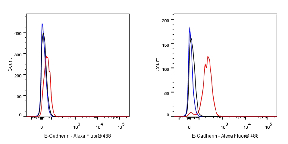
Flow cytometric analysis of 4% PFA fixed 90% methanol permeabilized MDA-MB-231 (Human breast adenocarcinoma epithelial cell, left) / HT29 (Human colorectal adenocarcinoma epithelial cell, Right) labelling E-Cadherin antibody at 1/50 dilution (1 μg) / (Red) compared with a Rabbit monoclonal IgG (Black) isotype control and an unlabelled control (cells without incubation with primary antibody and secondary antibody) (Blue). Goat Anti - Rabbit IgG Alexa Fluor 488® was used as the secondary antibody.
Negative control: MDA-MB-231
IHC shows positive staining in paraffin-embedded human liver. Anti-E-Cadherin antibody was used at 1/1000 dilution, followed by a HRP Polymer for Mouse & Rabbit IgG (ready to use). Counterstained with hematoxylin. Heat mediated antigen retrieval with Tris/EDTA buffer pH9.0 was performed before commencing with IHC staining protocol.
IHC shows positive staining in paraffin-embedded human kidney. Anti-E-Cadherin antibody was used at 1/1000 dilution, followed by a HRP Polymer for Mouse & Rabbit IgG (ready to use). Counterstained with hematoxylin. Heat mediated antigen retrieval with Tris/EDTA buffer pH9.0 was performed before commencing with IHC staining protocol.
IHC shows positive staining in paraffin-embedded human stomach. Anti-E-Cadherin antibody was used at 1/1000 dilution, followed by a HRP Polymer for Mouse & Rabbit IgG (ready to use). Counterstained with hematoxylin. Heat mediated antigen retrieval with Tris/EDTA buffer pH9.0 was performed before commencing with IHC staining protocol.
IHC shows positive staining in paraffin-embedded human breast cancer. Anti-E-Cadherin antibody was used at 1/1000 dilution, followed by a HRP Polymer for Mouse & Rabbit IgG (ready to use). Counterstained with hematoxylin. Heat mediated antigen retrieval with Tris/EDTA buffer pH9.0 was performed before commencing with IHC staining protocol.
IHC shows positive staining in paraffin-embedded human colon cancer. Anti-E-Cadherin antibody was used at 1/1000 dilution, followed by a HRP Polymer for Mouse & Rabbit IgG (ready to use). Counterstained with hematoxylin. Heat mediated antigen retrieval with Tris/EDTA buffer pH9.0 was performed before commencing with IHC staining protocol.
IHC shows positive staining in paraffin-embedded human endometrial cancer. Anti-E-Cadherin antibody was used at 1/1000 dilution, followed by a HRP Polymer for Mouse & Rabbit IgG (ready to use). Counterstained with hematoxylin. Heat mediated antigen retrieval with Tris/EDTA buffer pH9.0 was performed before commencing with IHC staining protocol.
IHC shows positive staining in paraffin-embedded human hepatocellular carcinoma. Anti-E-Cadherin antibody was used at 1/1000 dilution, followed by a HRP Polymer for Mouse & Rabbit IgG (ready to use). Counterstained with hematoxylin. Heat mediated antigen retrieval with Tris/EDTA buffer pH9.0 was performed before commencing with IHC staining protocol.
IHC shows positive staining in paraffin-embedded mouse colon. Anti-E-Cadherin antibody was used at 1/1000 dilution, followed by a HRP Polymer for Mouse & Rabbit IgG (ready to use). Counterstained with hematoxylin. Heat mediated antigen retrieval with Tris/EDTA buffer pH9.0 was performed before commencing with IHC staining protocol.
IHC shows positive staining in paraffin-embedded rat kidney. Anti-E-Cadherin antibody was used at 1/1000 dilution, followed by a HRP Polymer for Mouse & Rabbit IgG (ready to use). Counterstained with hematoxylin. Heat mediated antigen retrieval with Tris/EDTA buffer pH9.0 was performed before commencing with IHC staining protocol.
ICC shows positive staining in HT-29 cells. Anti-E-Cadherin antibody was used at 1/500 dilution (Green) and incubated overnight at 4°C. Goat polyclonal Antibody to Rabbit IgG - H&L (Alexa Fluor® 488) was used as secondary antibody at 1/1000 dilution. The cells were fixed with 4% PFA and permeabilized with 0.1% PBS-Triton X-100. Nuclei were counterstained with DAPI (Blue).
ICC shows positive staining in MCF7 cells. Anti-E-Cadherin antibody was used at 1/500 dilution (Green) and incubated overnight at 4°C. Goat polyclonal Antibody to Rabbit IgG - H&L (Alexa Fluor® 488) was used as secondary antibody at 1/1000 dilution. The cells were fixed with 4% PAF and permeabilized with 0.1% PBS-Triton X-100. Nuclei were counterstained with DAPI (Blue).
Negative control: ICC shows negative staining in MDA-MB-231 cells. Anti-E-Cadherin antibody was used at 1/500 dilution and incubated overnight at 4°C. Goat polyclonal Antibody to Rabbit IgG - H&L (Alexa Fluor® 488) was used as secondary antibody at 1/1000 dilution. The cells were fixed with 4% PFA and permeabilized with 0.1% PBS-Triton X-100. Nuclei were counterstained with DAPI (Blue).
 您现在的位置: