
12 months from date of receipt / reconstitution, -20 °C as supplied
| 应用 | 稀释度 |
|---|---|
| WB | 1:1000 |
| FC | 1:500 |
| ICC | 1:100 |
| IHC-P | 1:100 |
SMAD (mothers against decapentaplegic homologs) molecules are the core components in TGF-β signaling pathway. TGF-β binding to its receptor induces phosphorylation and activation of receptor-regulated SMADs (R-SMADs), SMAD2 and SMAD3, which subsequently associate with their partner SMAD4 and translocate from cytoplasm to nucleus. Formation of R-SMAD–SMAD4 complexes is essential in signaling of most TGF-β family members.
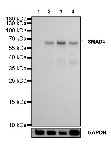
WB result of SMAD4 Rabbit mAb Primary antibody: SMAD4 Rabbit mAb at 1/1000 dilution Lane 1: HT-29 whole cell lysate 20 µg Lane 2: HepG2 whole cell lysate 20 µg Lane 3: HCT 116 whole cell lysate 20 µg Lane 4: Jurkat whole cell lysate 20 µg Negative control: HT-29 whole cell lysate Secondary antibody: Goat Anti-Rabbit IgG, (H+L), HRP conjugated at 1/10000 dilution Predicted MW: 60 kDa Observed MW: 65 kDa
Flow cytometric analysis of HT-29 (Human colorectal adenocarcinoma epithelial cell, left) / HepG2 (Human hepatocellular carcinoma epithelial cell, right) cells labelling SMAD4 antibody at 1/500 dilution (0.1 μg)/ (red) compared with a Rabbit monoclonal IgG (Black) isotype control and an unlabelled control (cells without incubation with primary antibody and secondary antibody) (Blue). Goat Anti-Rabbit IgG Alexa Fluor® 488 was used as the secondary antibody. Negative control: HT-29 cells
IHC shows positive staining in paraffin-embedded human colon cancer. Anti-SMAD4 antibody was used at 1/100 dilution, followed by a HRP Polymer for Mouse & Rabbit IgG (ready to use). Counterstained with hematoxylin. Heat mediated antigen retrieval with Tris/EDTA buffer pH9.0 was performed before commencing with IHC staining protocol.
IHC shows positive staining in paraffin-embedded human colon cancer. Anti-SMAD4 antibody was used at 1/100 dilution, followed by a HRP Polymer for Mouse & Rabbit IgG (ready to use). Counterstained with hematoxylin. Heat mediated antigen retrieval with Tris/EDTA buffer pH9.0 was performed before commencing with IHC staining protocol.
IHC shows positive staining in paraffin-embedded human lung squamous cell carcinoma. Anti-SMAD4 antibody was used at 1/100 dilution, followed by a HRP Polymer for Mouse & Rabbit IgG (ready to use). Counterstained with hematoxylin. Heat mediated antigen retrieval with Tris/EDTA buffer pH9.0 was performed before commencing with IHC staining protocol.
IHC shows positive staining in paraffin-embedded human pancreatic cancer (Loss of SMAD4 expression in tumors but nuclear expression in stromal cells). Anti-SMAD4 antibody was used at 1/100 dilution, followed by a HRP Polymer for Mouse & Rabbit IgG (ready to use). Counterstained with hematoxylin. Heat mediated antigen retrieval with Tris/EDTA buffer pH9.0 was performed before commencing with IHC staining protocol.
IHC shows positive staining in paraffin-embedded human pancreatic cancer (Loss of SMAD4 expression in tumors but expression in stromal and paracancerous cells). Anti-SMAD4 antibody was used at 1/100 dilution, followed by a HRP Polymer for Mouse & Rabbit IgG (ready to use). Counterstained with hematoxylin. Heat mediated antigen retrieval with Tris/EDTA buffer pH9.0 was performed before commencing with IHC staining protocol.
ICC shows positive staining in HepG2 cells. Anti-SMAD4 antibody was used at 1/100 dilution (Green) and incubated overnight at 4°C. Goat polyclonal Antibody to Rabbit IgG - H&L (Alexa Fluor® 488) was used as secondary antibody at 1/1000 dilution. The cells were fixed with 4% PFA and permeabilized with 0.1% PBS-Triton X-100. Nuclei were counterstained with DAPI (Blue).
Negative control: ICC shows negative staining in HT-29 cells. Anti-SMAD4 antibody was used at 1/100 dilution and incubated overnight at 4°C. Goat polyclonal Antibody to Rabbit IgG - H&L (Alexa Fluor® 488) was used as secondary antibody at 1/1000 dilution. The cells were fixed with 4% PFA and permeabilized with 0.1% PBS-Triton X-100. Nuclei were counterstained with DAPI (Blue).
 您现在的位置: