
PBS, 40% Glycerol, 0.05% BSA, 0.03% Proclin 300
12 months from date of receipt / reconstitution, -20 °C as supplied.
| 应用 | 稀释度 |
|---|---|
| WB | 1:1000 |
| IHC | 1:500-1:1000 |
| ICC | 1:1000 |
| IF | 1:500 |
| ICFCM | 1:500 |
Catenin δ-1 (P120 Catenin) belongs to the β-serial protein family .p-120 catenin (along with α, β and γ catenins) connects the transmembrane protein E-cadherin to the actin cytoskeleton in the cell cytoplasm. It expresses in various normal epithelial tissues (including thyroid, breast, stomach, duodenal, liver, pancreas, colon, kidney, prostate, testicles), various non -epithelial tissues (including myocardial cells, neurons, gel cells To. p-120 regulates the transformation of calcium adhesion protein on the surface of the cells, thereby determining the E-calcium adhesive protein levels used in the cells, and then plays an important role in the adhesion of the cells. Many studies have shown that p-120 irregularly expression or lack of disobedience in a variety of cancer cells indicates that p-120 play a role in cancer-suppressing genes.
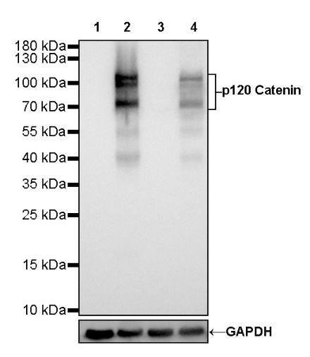
WB result of p120 Catenin Rabbit mAb
Primary antibody: p120 Catenin Rabbit mAb at 1/1000 dilution
Lane 1: HL-60 whole cell lysate 20 µg
Lane 2: HT-29 whole cell lysate 20 µg
Lane 3: HeLa whole cell lysate 20 µg
Lane 4: A431 whole cell lysate 20 µg
Negative control: HL-60 whole cell lysate
Secondary antibody: Goat Anti-Rabbit IgG, (H+L), HRP conjugated at 1/10000 dilution
Predicted MW: 108 kDa
Observed MW: 70~105 kDa
Exposure time: 3 s
Flow cytometric analysis of 4% PFA fixed 90% methanol permeabilized HL-60 (Human acute promyelocytic leukemia promyeloblast, left) / HT29 (Human colorectal adenocarcinoma epithelial cell, Right) cells labelling E-Cadherin antibody at 1/500 dilution (0.1 μg) / (red) compared with a Rabbit monoclonal IgG (Black) isotype control and an unlabelled control (cells without incubation with primary antibody and secondary antibody) (Blue). Goat Anti - Rabbit IgG Alexa Fluor® 488 was used as the secondary antibody. Negative control: HL-60
IHC shows positive staining in paraffin-embedded human tonsil. Anti-p120 Catenin antibody was used at 1/500 dilution, followed by a HRP Polymer for Mouse & Rabbit IgG (ready to use). Counterstained with hematoxylin. Heat mediated antigen retrieval with Tris/EDTA buffer pH9.0 was performed before commencing with IHC staining protocol.
IHC shows positive staining in paraffin-embedded human stomach. Anti-p120 Catenin antibody was used at 1/500 dilution, followed by a HRP Polymer for Mouse & Rabbit IgG (ready to use). Counterstained with hematoxylin. Heat mediated antigen retrieval with Tris/EDTA buffer pH9.0 was performed before commencing with IHC staining protocol.
IHC shows positive staining in paraffin-embedded human breast cancer. Anti-p120 Catenin antibody was used at 1/500 dilution, followed by a HRP Polymer for Mouse & Rabbit IgG (ready to use). Counterstained with hematoxylin. Heat mediated antigen retrieval with Tris/EDTA buffer pH9.0 was performed before commencing with IHC staining protocol.
IHC shows positive staining in paraffin-embedded human colon cancer. Anti-p120 Catenin antibody was used at 1/500 dilution, followed by a HRP Polymer for Mouse & Rabbit IgG (ready to use). Counterstained with hematoxylin. Heat mediated antigen retrieval with Tris/EDTA buffer pH9.0 was performed before commencing with IHC staining protocol.
IHC shows positive staining in paraffin-embedded mouse stomach. Anti-p120 Catenin antibody was used at 1/500 dilution, followed by a HRP Polymer for Mouse & Rabbit IgG (ready to use). Counterstained with hematoxylin. Heat mediated antigen retrieval with Tris/EDTA buffer pH9.0 was performed before commencing with IHC staining protocol.
IHC shows positive staining in paraffin-embedded rat liver. Anti-p120 Catenin antibody was used at 1/500 dilution, followed by a HRP Polymer for Mouse & Rabbit IgG (ready to use). Counterstained with hematoxylin. Heat mediated antigen retrieval with Tris/EDTA buffer pH9.0 was performed before commencing with IHC staining protocol.
IHC shows positive staining in paraffin-embedded rat esophagus. Anti-p120 Catenin antibody was used at 1/500 dilution, followed by a HRP Polymer for Mouse & Rabbit IgG (ready to use). Counterstained with hematoxylin. Heat mediated antigen retrieval with Tris/EDTA buffer pH9.0 was performed before commencing with IHC staining protocol.
ICC shows positive staining in HT-29 cells. Anti-p120 Catenin antibody was used at 1/1000 dilution (Green) and incubated overnight at 4°C. Goat polyclonal Antibody to Rabbit IgG - H&L (Alexa Fluor® 488) was used as secondary antibody at 1/1000 dilution. The cells were fixed with 4% PFA and permeabilized with 0.1% PBS-Triton X-100. Nuclei were counterstained with DAPI (Blue).
Negative control: ICC shows negative staining in HL-60 cells. Anti-p120 Catenin antibody was used at 1/1000 dilution (Green) and incubated overnight at 4°C. Goat polyclonal Antibody to Rabbit IgG - H&L (Alexa Fluor® 488) was used as secondary antibody at 1/1000 dilution. The cells were fixed with 4% PFA and permeabilized with 0.1% PBS-Triton X-100. Nuclei were counterstained with DAPI (Blue).
IF shows positive staining in paraffin-embedded human gastric cancer. Anti- p120 Catenin antibody was used at 1/500 dilution (Green) and incubated overnight at 4°C. Goat polyclonal Antibody to Rabbit IgG - H&L (Alexa Fluor® 488) (S0B4004) was used as secondary antibody at 1/1000 dilution. Counterstained with DAPI (Blue). Heat mediated antigen retrieval with EDTA buffer pH9.0 was performed before commencing with IF staining protocol.
IF shows positive staining in paraffin-embedded human colon cancer. Anti- p120 Catenin antibody was used at 1/500 dilution (Green) and incubated overnight at 4°C. Goat polyclonal Antibody to Rabbit IgG - H&L (Alexa Fluor® 488) (S0B4004) was used as secondary antibody at 1/1000 dilution. Counterstained with DAPI (Blue). Heat mediated antigen retrieval with EDTA buffer pH9.0 was performed before commencing with IF staining protocol.
 您现在的位置: