
12 months from date of receipt / reconstitution, -20 °C as supplied
| 应用 | 稀释度 |
|---|---|
| WB | 1:1000 |
| ICC | 1:50 |
DNA mismatch repair protein Mlh1 or MutL protein homolog 1 is a protein that in humans is encoded by the MLH1 gene located on chromosome 3. It is a gene commonly associated with hereditary nonpolyposis colorectal cancer. MLH1 protein is one component of a system of seven DNA mismatch repair proteins that work coordinately in sequential steps to initiate repair of DNA mismatches in humans [PMID: 18543306]. Defects in mismatch repair, found in about 13% of colorectal cancers, are much more frequently due to deficiency of MLH1 than deficiencies of other DNA mismatch repair proteins [PMID: 15887099]. The seven DNA mismatch repair proteins in humans are MLH1, MLH3, MSH2, MSH3, MSH6, PMS1 and PMS2 [ PMID: 18543306].

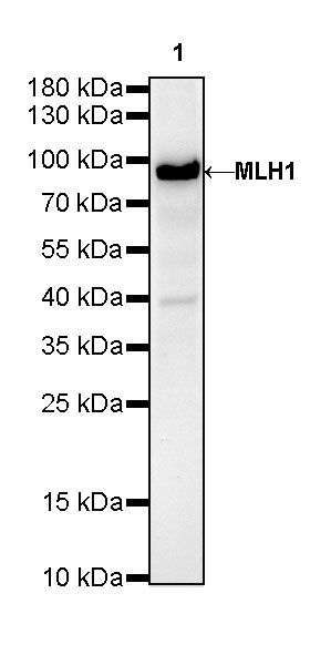
WB result of MLH1 Rabbit mAb
Primary antibody: MLH1 Rabbit mAb at 1/1000 dilution
Lane 1: rat testis lysate 20 µg
Secondary antibody: Goat Anti-Rabbit IgG, (H+L), HRP conjugated at 1/10000 dilution
Predicted MW: 84 kDa
Observed MW: 84 kDa
(This blot was developed with high sensitivity substrate)
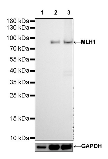
WB result of MLH1 Rabbit mAb
Primary antibody: MLH1 Rabbit mAb at 1/1000 dilution
Lane 1: HCT 116 whole cell lysate 20 µg
Lane 2: HeLa whole cell lysate 20 µg
Lane 3: Jurkat whole cell lysate 20 µg
Negative control: HCT 116 whole cell lysate
Secondary antibody: Goat Anti-Rabbit IgG, (H+L), HRP conjugated at 1/10000 dilution
Predicted MW: 84 kDa
Observed MW: 84 kDa
ICC shows positive staining in HeLa cells. Anti-MLH1 antibody was used at 1/50 dilution (Green) and incubated overnight at 4°C. Goat polyclonal Antibody to Rabbit IgG - H&L (Alexa Fluor® 488) was used as secondary antibody at 1/1000 dilution. The cells were fixed with 4% PFA and permeabilized with 0.1% PBS-Triton X-100. Nuclei were counterstained with DAPI (Blue).
Negative control: ICC shows negative staining in HCT-116 cells. Anti-MLH1 antibody was used at 1/50 dilution (Green) and incubated overnight at 4°C. Goat polyclonal Antibody to Rabbit IgG - H&L (Alexa Fluor® 488) was used as secondary antibody at 1/1000 dilution. The cells were fixed with 4% PFA and permeabilized with 0.1% PBS-Triton X-100. Nuclei were counterstained with DAPI (Blue).
 您现在的位置: