
12 months from date of receipt / reconstitution, -20 °C as supplied
| 应用 | 稀释度 |
|---|---|
| WB | 1:1000 |
| IHC-P | 1:500 |
| ICC | 1:200-1:500 |
| IP | 1:50 |
FOXA2 is member of a large family of nuclear transcription factors, termed the winged helix family of transcription factors, that are involved in cell commitment, differentiation, and gene transcription in a variety of organs, such as the central nervous system and derivatives of the foregut endoderm, including the gastrointestinal tract, lung, and liver. FOXA2 is required for the formation of foregut endoderm, from which the lung bud is derived, and plays a critical role in organogenesis of the lung.

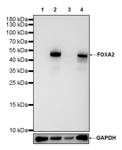
WB result of FOXA2 Rabbit mAb
Primary antibody: FOXA2 Rabbit mAb at 1/1000 dilution
Lane 1: LNCaP whole cell lysate 20 µg
Lane 2: PC-3 whole cell lysate 20 µg
Lane 3: SW480 whole cell lysate 20 µg
Lane 4: HepG2 whole cell lysate 20 µg
Negative control: LNCaP whole cell lysate
Secondary antibody: Goat Anti-Rabbit IgG, (H+L), HRP conjugated at 1/10000 dilution
Predicted MW: 48kDa
Observed MW: 48kDa

Flow cytometric analysis of HeLa (Human cervix adenocarcinoma epithelial cell, left) / HepG2 (Human hepatocellular carcinoma epithelial cell, right) cells labelling FOXA2 antibody at 1/500 dilution (0.1 μg)/ (red) compared with a Rabbit monoclonal IgG (Black) isotype control and an unlabelled control (cells without incubation with primary antibody and secondary antibody) (Blue). Goat Anti-Rabbit IgG Alexa Fluor® 488 was used as the secondary antibody.
Negative control: HeLa
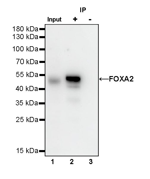
FOXA2 Rabbit mAb at 1/50 dilution (1 µg) immunoprecipitating FOXA2 in 0.4 mg PC-3 whole cell lysate.
Western blot was performed on the immunoprecipitate using FOXA2 Rabbit mAb at 1/1000 dilution.
Secondary antibody (HRP) for IP was used at 1/400 dilution.
Lane 1: PC-3 whole cell lysate 20 µg (Input)
Lane 2: FOXA2 Rabbit mAb IP in PC-3 whole cell lysate
Lane 3: Rabbit monoclonal IgG IP in PC-3 whole cell lysate
Predicted MW: 48 kDa
Observed MW: 48 kDa
(This blot was developed with high sensitivity substrate)

IHC shows positive staining in paraffin-embedded human stomach. Anti-FOXA2 antibody was used at 1/500 dilution, followed by a HRP Polymer for Mouse & Rabbit IgG (ready to use). Counterstained with hematoxylin. Heat mediated antigen retrieval with Tris/EDTA buffer pH9.0 was performed before commencing with IHC staining protocol.
(Weak affinity compared to mice)
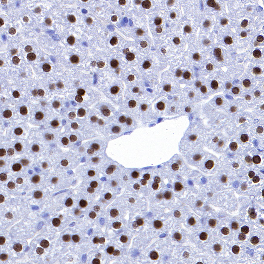
IHC shows positive staining in paraffin-embedded mouse liver. Anti-FOXA2 antibody was used at 1/500 dilution, followed by a HRP Polymer for Mouse & Rabbit IgG (ready to use). Counterstained with hematoxylin. Heat mediated antigen retrieval with Tris/EDTA buffer pH9.0 was performed before commencing with IHC staining protocol.

IHC shows positive staining in paraffin-embedded mouse lung. Anti-FOXA2 antibody was used at 1/500 dilution, followed by a HRP Polymer for Mouse & Rabbit IgG (ready to use). Counterstained with hematoxylin. Heat mediated antigen retrieval with Tris/EDTA buffer pH9.0 was performed before commencing with IHC staining protocol.

IHC shows positive staining in paraffin-embedded mouse stomach. Anti-FOXA2 antibody was used at 1/500 dilution, followed by a HRP Polymer for Mouse & Rabbit IgG (ready to use). Counterstained with hematoxylin. Heat mediated antigen retrieval with Tris/EDTA buffer pH9.0 was performed before commencing with IHC staining protocol.

IHC shows positive staining in paraffin-embedded rat liver. Anti-FOXA2 antibody was used at 1/200 dilution, followed by a HRP Polymer for Mouse & Rabbit IgG (ready to use). Counterstained with hematoxylin. Heat mediated antigen retrieval with Tris/EDTA buffer pH9.0 was performed before commencing with IHC staining protocol.

IHC shows positive staining in paraffin-embedded rat lung. Anti-FOXA2 antibody was used at 1/500 dilution, followed by a HRP Polymer for Mouse & Rabbit IgG (ready to use). Counterstained with hematoxylin. Heat mediated antigen retrieval with Tris/EDTA buffer pH9.0 was performed before commencing with IHC staining protocol.

IHC shows positive staining in paraffin-embedded rat stomach. Anti-FOXA2 antibody was used at 1/500 dilution, followed by a HRP Polymer for Mouse & Rabbit IgG (ready to use). Counterstained with hematoxylin. Heat mediated antigen retrieval with Tris/EDTA buffer pH9.0 was performed before commencing with IHC staining protocol.
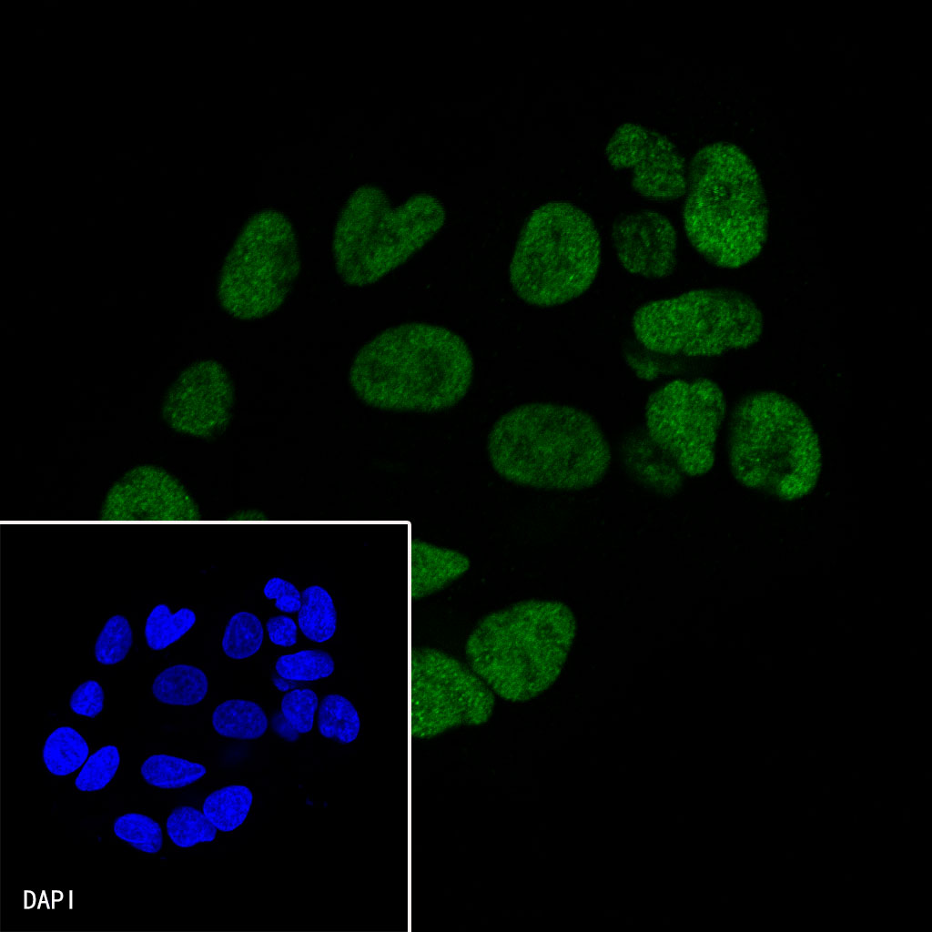
ICC shows positive staining in HepG2 cells. Anti-FOXA2 antibody was used at 1/500 dilution and incubated overnight at 4°C. Goat polyclonal Antibody to Rabbit IgG - H&L (Alexa Fluor® 488) was used as secondary antibody at 1/1000 dilution.The cells were fixed with 4% FPA and permeabilized with 0.1% PBS-Triton X-100. Nuclei were counterstained with DAPI.

Negative control: ICC shows negative staining in HeLa cells. Anti-FOXA2 antibody was used at 1/500 dilution and incubated overnight at 4°C. Goat polyclonal Antibody to Rabbit IgG - H&L (Alexa Fluor® 488) was used as secondary antibody at 1/1000 dilution.The cells were fixed with 4% FPA and permeabilized with 0.1% PBS-Triton X-100. Nuclei were counterstained with DAPI.
 您现在的位置: