
12 months from date of receipt / reconstitution, -20 °C as supplied
| 应用 | 稀释度 |
|---|---|
| WB | 1:1250 |
| IHC-P | 1:1000 |
| ICC | 1:125 |
| ICFCM | 1:500 |
CD45 is a type I transmembrane protein that is present in various isoforms on all differentiated hematopoietic cells (except erythrocytes and plasma cells). CD45 has been shown to be an essential regulator of T- and B-cell antigen receptor signaling. It functions through either direct interaction with components of the antigen receptor complexes via its extracellular domain (a form of co-stimulation), or by activating various Src family kinases required for the antigen receptor signaling via its cytoplasmic domain. CD45 also suppresses JAK kinases, and so functions as a negative regulator of cytokine receptor signaling.
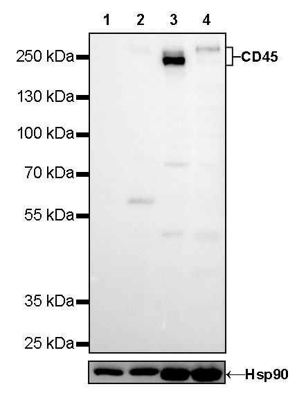
WB result of CD45 Mouse mAb
Primary antibody: CD45 Mouse mAb at 1/1250 dilution
Lane 1: MCF7 whole cell lysate 20 µg
Lane 2: THP-1 whole cell lysate 20 µg
Lane 3: Jurkat whole cell lysate 20 µg
Lane 4: Raji whole cell lysate 20 µg
Negative control: MCF7 whole cell lysate
Low expression control: THP-1 whole cell lysate
Secondary antibody: Goat Anti-Mouse IgG, (H+L), HRP conjugated at 1/10000 dilution
Predicted MW: 147kDa
Observed MW: 240~260kDa

Flow cytometric analysis of 4% PFA fixed 90% methanol permeabilized Jurkat (Human T cell leukemia T lymphocyte) cells labelling Amyloid Precursor Protein antibody at 1/500 dilution (0.1 μg)/ (Red) compared with a Mouse monoclonal IgG (Black) isotype control and an unlabelled control (cells without incubation with primary antibody and secondary antibody) (Blue). Goat Anti - Mouse IgG Alexa Fluor® 488 was used as the secondary antibody.

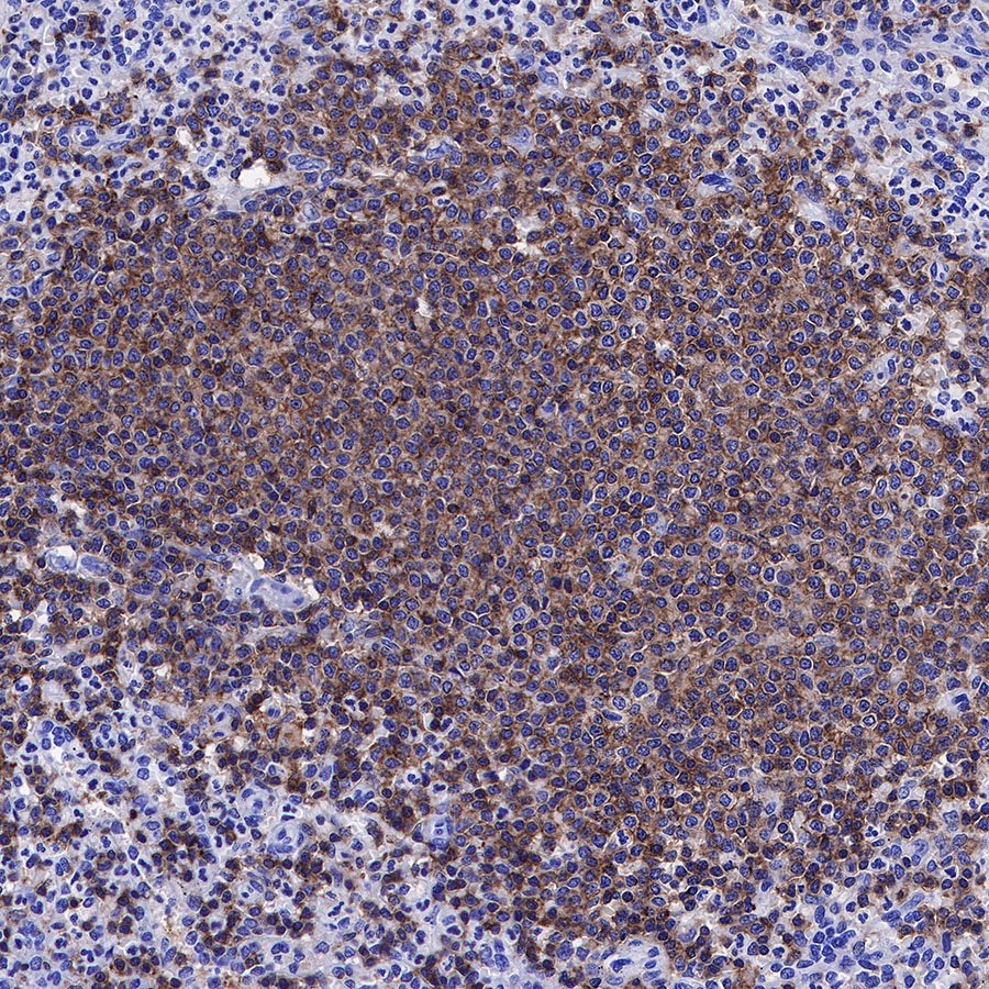
IHC shows positive staining in paraffin-embedded human tonsil. Anti-CD45 antibody was used at 1/1000 dilution, followed by a HRP Polymer for Mouse & Rabbit IgG (ready to use). Counterstained with hematoxylin. Heat mediated antigen retrieval with Tris/EDTA buffer pH9.0 was performed before commencing with IHC staining protocol.
IHC shows positive staining in paraffin-embedded human spleen. Anti-CD45 antibody was used at 1/1000 dilution, followed by a HRP Polymer for Mouse & Rabbit IgG (ready to use). Counterstained with hematoxylin. Heat mediated antigen retrieval with Tris/EDTA buffer pH9.0 was performed before commencing with IHC staining protocol.

IHC shows positive staining in paraffin-embedded human colon. Anti-CD45 antibody was used at 1/1000 dilution, followed by a HRP Polymer for Mouse & Rabbit IgG (ready to use). Counterstained with hematoxylin. Heat mediated antigen retrieval with Tris/EDTA buffer pH9.0 was performed before commencing with IHC staining protocol.
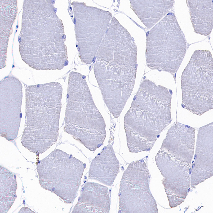
Negative control: IHC shows negative staining in paraffin-embedded human skeletal muscle. Anti-CD45 antibody was used at 1/1000 dilution, followed by a HRP Polymer for Mouse & Rabbit IgG (ready to use). Counterstained with hematoxylin. Heat mediated antigen retrieval with Tris/EDTA buffer pH9.0 was performed before commencing with IHC staining protocol.
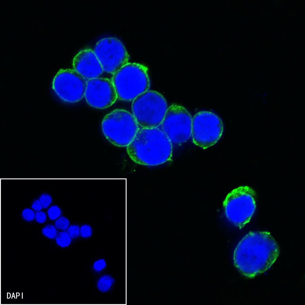
ICC shows positive staining in Jurkat cells. Anti-CD45 antibody was used at 1/125 dilution (Green) and incubated overnight at 4°C. Goat polyclonal Antibody to mouse IgG - H&L (Alexa Fluor® 488) was used as secondary antibody at 1/1000 dilution. The cells were fixed with 100% ice-cold methanol and permeabilized with 0.1% PBS-Triton X-100. Nuclei were counterstained with DAPI (Blue).
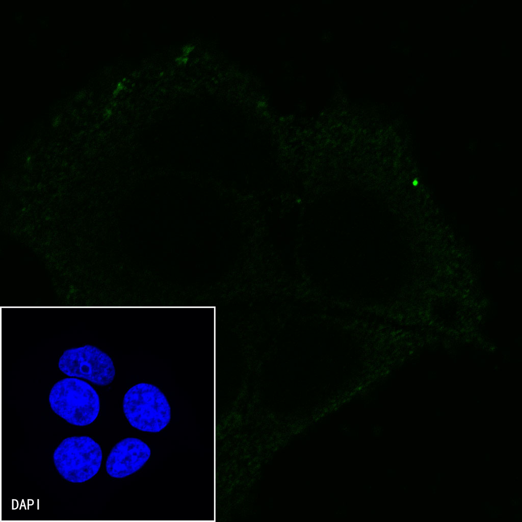
Negative control:ICC shows negative staining in MCF7 cells.Anti-CD45 antibody was used at 1/125 dilution and incubated overnight at 4°C. Goat polyclonal Antibody to mouse IgG - H&L (Alexa Fluor® 488) was used as secondary antibody at 1/1000 dilution. The cells were fixed with 100% ice-cold methanol and permeabilized with 0.1% PBS-Triton X-100. Nuclei were counterstained with DAPI (Blue).
 您现在的位置:
