

12 months from date of receipt / reconstitution, -20 °C as supplied
| 应用 | 稀释度 |
|---|---|
| WB | 1:1000 |
| IHC-P | 1:500 |
The cell adhesion molecule (CAM), N-cadherin, has emerged as an important oncology therapeutic target. N-cadherin is a transmembrane glycoprotein mediating the formation and structural integrity of blood vessels. Its expression has also been documented in numerous types of poorly differentiated tumours [PMID: 25533096].
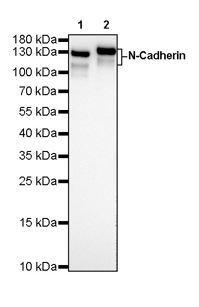
WB result of N-Cadherin Rabbit mAb
Primary antibody: N-Cadherin Rabbit mAb at 1/1000 dilution
Lane 1: rat brain lysate 20 µg
Lane 2: rat heart lysate 20 µg
Secondary antibody: Goat Anti-Rabbit IgG, (H+L), HRP conjugated at 1/10000 dilution
Predicted MW: 100kDa
Observed MW: 110~140kDa
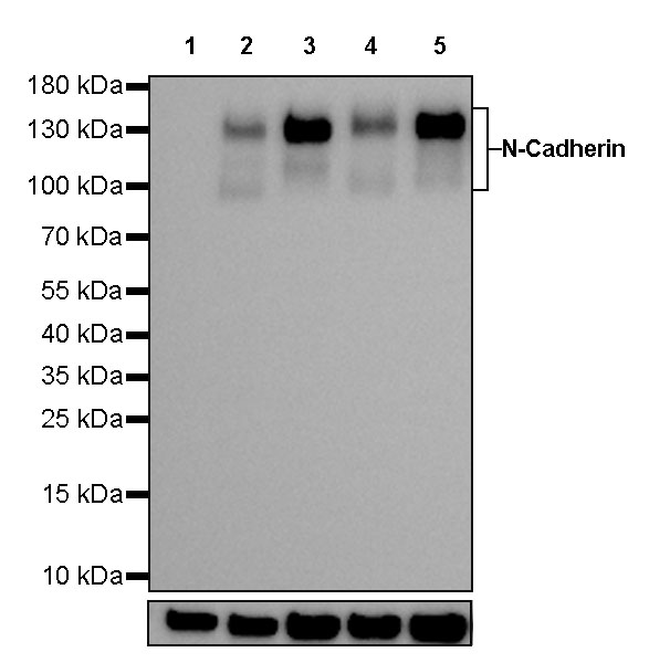
WB result of N-Cadherin Rabbit mAb
Primary antibody: N-Cadherin Rabbit mAb at 1/1000 dilution
Lane 1: MCF7 whole cell lysate 20 µg
Lane 2: A549 whole cell lysate 20 µg
Lane 3: HeLa whole cell lysate 20 µg
Lane 4: PC-3 whole cell lysate 20 µg
Lane 5: HepG2 whole cell lysate 20 µg
Negative control: MCF7 whole cell lysate
Secondary antibody: Goat Anti-Rabbit IgG, (H+L), HRP conjugated at 1/10000 dilution
Predicted MW: 100kDa
Observed MW: 110~140kDa
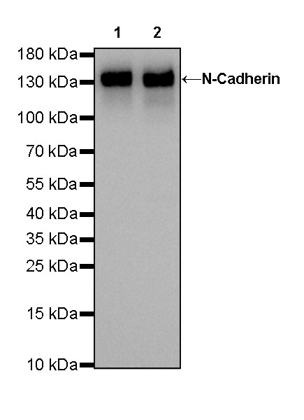
WB result of N-Cadherin Rabbit mAb
Primary antibody: N-Cadherin Rabbit mAb at 1/1000 dilution
Lane 1: mouse brain lysate 20 µg
Lane 2: mouse heart lysate 20 µg
Secondary antibody: Goat Anti-Rabbit IgG, (H+L), HRP conjugated at 1/10000 dilution
Predicted MW: 100kDa
Observed MW: 140kDa
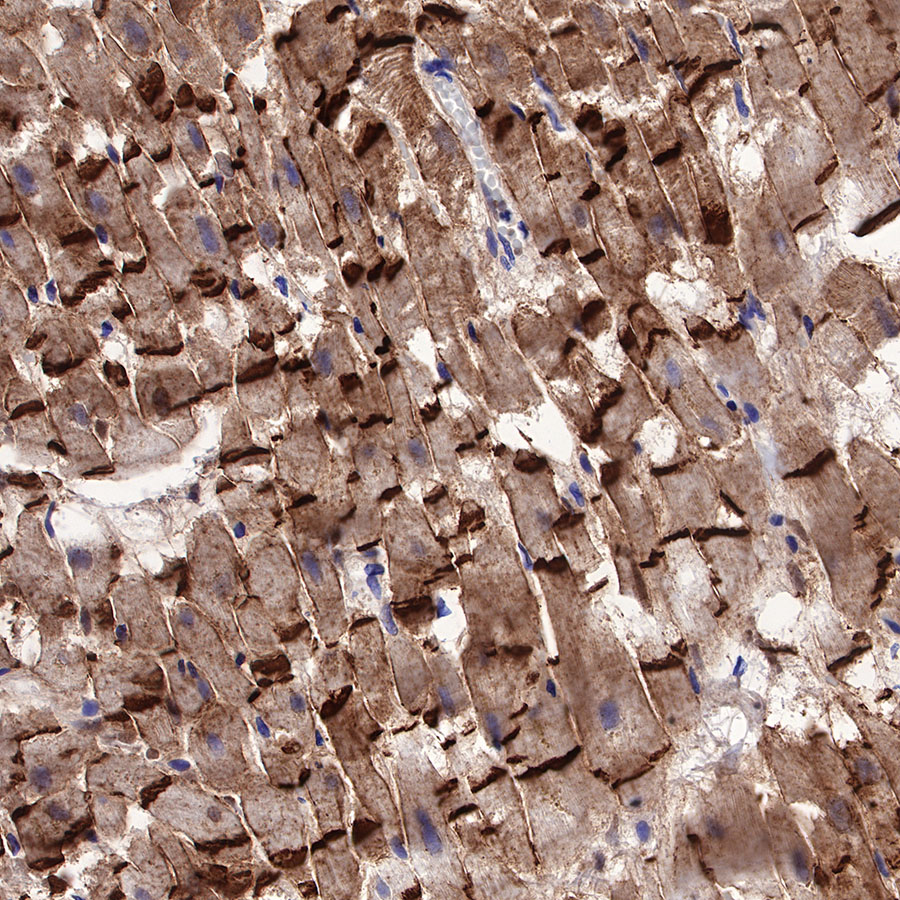
IHC shows positive staining in paraffin-embedded human cardiac muscle. Anti-N-Cadherin antibody was used at 1/500 dilution, followed by a HRP Polymer for Mouse & Rabbit IgG (ready to use). Counterstained with hematoxylin. Heat mediated antigen retrieval with Tris/EDTA buffer pH9.0 was performed before commencing with IHC staining protocol.
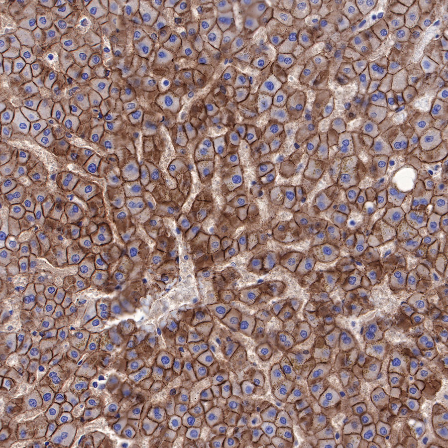
IHC shows positive staining in paraffin-embedded human liver. Anti-N-Cadherin antibody was used at 1/500 dilution, followed by a HRP Polymer for Mouse & Rabbit IgG (ready to use). Counterstained with hematoxylin. Heat mediated antigen retrieval with Tris/EDTA buffer pH9.0 was performed before commencing with IHC staining protocol.
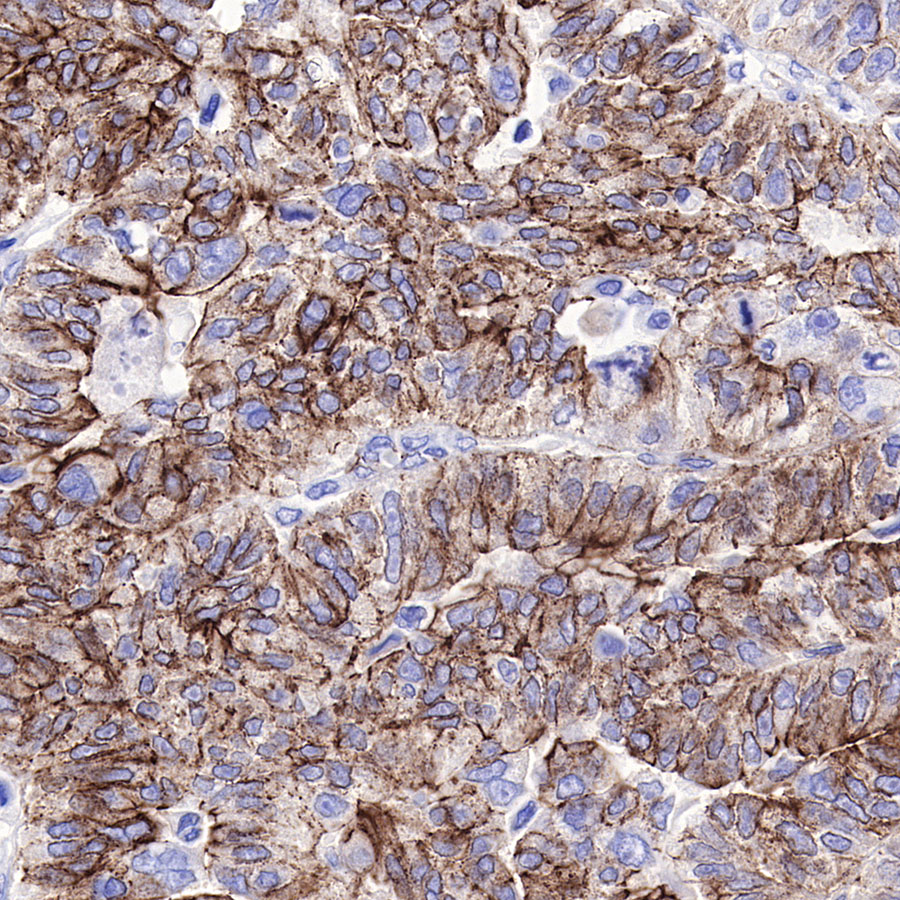
IHC shows positive staining in paraffin-embedded human ovarian carcinoma. Anti-N-Cadherin antibody was used at 1/500 dilution, followed by a HRP Polymer for Mouse & Rabbit IgG (ready to use). Counterstained with hematoxylin. Heat mediated antigen retrieval with Tris/EDTA buffer pH9.0 was performed before commencing with IHC staining protocol.
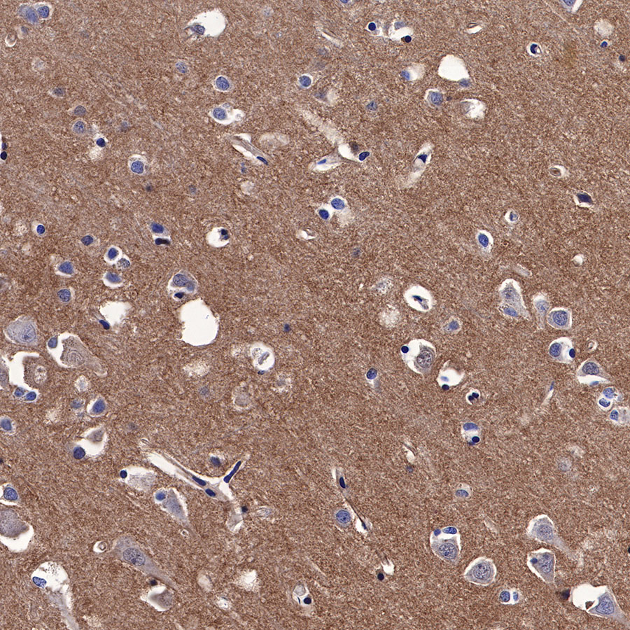
IHC shows positive staining in paraffin-embedded human cerebral cortex. Anti-N-Cadherin antibody was used at 1/500 dilution, followed by a HRP Polymer for Mouse & Rabbit IgG (ready to use). Counterstained with hematoxylin. Heat mediated antigen retrieval with Tris/EDTA buffer pH9.0 was performed before commencing with IHC staining protocol.
IHC shows positive staining in paraffin-embedded human renal clear cell carcinoma. Anti-N-Cadherin antibody was used at 1/500 dilution, followed by a HRP Polymer for Mouse & Rabbit IgG (ready to use). Counterstained with hematoxylin. Heat mediated antigen retrieval with Tris/EDTA buffer pH9.0 was performed before commencing with IHC staining protocol.
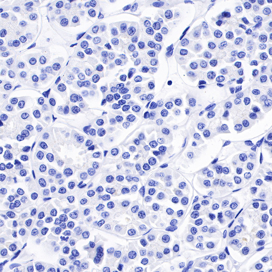
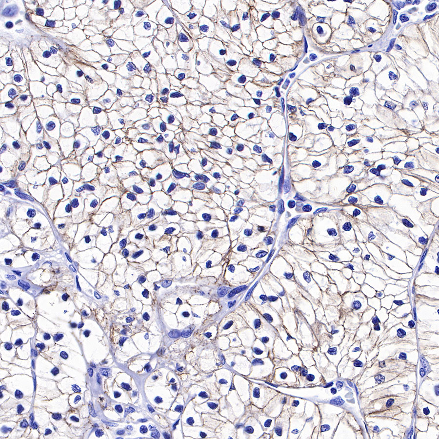
Negative tissue: IHC shows negative staining in paraffin-embedded human chromophobe renal carcinoma. Anti-N-Cadherin antibody was used at 1/500 dilution, followed by a HRP Polymer for Mouse & Rabbit IgG (ready to use). Counterstained with hematoxylin. Heat mediated antigen retrieval with Tris/EDTA buffer pH9.0 was performed before commencing with IHC staining protocol.
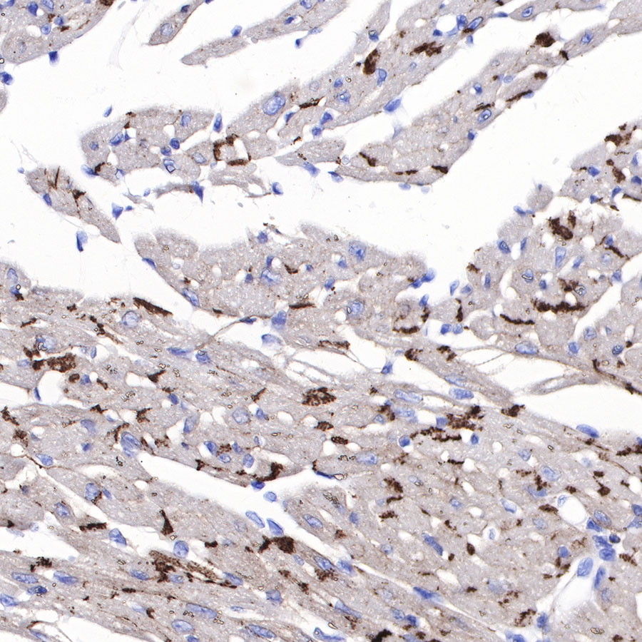
IHC shows positive staining in paraffin-embedded mouse cardiac muscle. Anti-N-Cadherin antibody was used at 1/500 dilution, followed by a HRP Polymer for Mouse & Rabbit IgG (ready to use). Counterstained with hematoxylin. Heat mediated antigen retrieval with Tris/EDTA buffer pH9.0 was performed before commencing with IHC staining protocol.
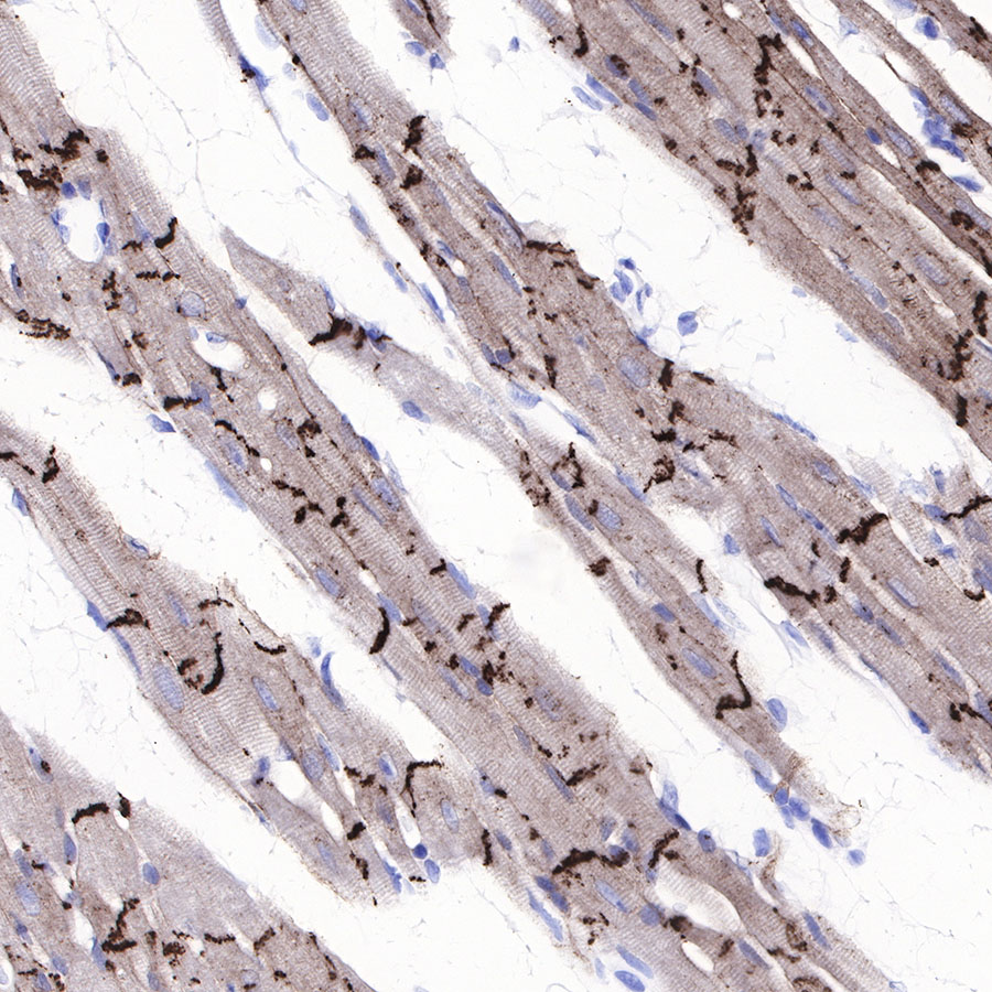
IHC shows positive staining in paraffin-embedded rat cardiac muscle. Anti-N-Cadherin antibody was used at 1/500 dilution, followed by a HRP Polymer for Mouse & Rabbit IgG (ready to use). Counterstained with hematoxylin. Heat mediated antigen retrieval with Tris/EDTA buffer pH9.0 was performed before commencing with IHC staining protocol.
 您现在的位置: