

12 months from date of receipt / reconstitution, -20 °C as supplied
| 应用 | 稀释度 |
|---|---|
| WB | 1:1000 |
| ICC | 1:250 |
Insulin-like growth factor binding proteins (IGFBPs) are a family of proteins binding to Insulin-like growth factors (IGFs), generally including IGFBP1, IGFBP2, IGFBP3, IGFBP4, IGFBP5, and IGFBP6 [PMID: 30214426]. IGFBP1, a 40 to 50 kDa protein, which plays an indispensable role in normal growth and development, and in the pathophysiology of various tumors. IGFBP-1 has been shown to be associated with the risk of various tumors, and has a vital function in regulating tumor behaviors such as proliferation, migration, invasion and adhesion through different molecular mechanisms. The biological actions of IGFBP-1 in cancer are found to be related to its phosphorylation state, and the IGF-dependent and -independent mechanisms. [PMID: 1648311, PMID: 33841624].
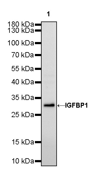
WB result of IGFBP1 Rabbit mAb
Primary antibody: IGFBP1 Rabbit mAb at 1/1000 dilution
Lane 1: HepG2 whole cell lysate 20 µg
Secondary antibody: Goat Anti-Rabbit IgG, (H+L), HRP conjugated at 1/10000 dilution
Predicted MW: 28 kDa
Observed MW: 30 kDa
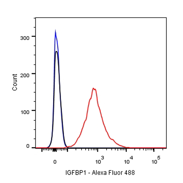
Flow cytometric analysis of HepG2 cells labelling IGFBP1 antibody at 1/500 (0.1 μg) dilution/ (red) compared with a Rabbit monoclonal IgG (Black) isotype control and an unlabelled control (cells without incubation with primary antibody and secondary antibody) (Blue). Goat Anti-Rabbit IgG Alexa Fluor® 488 was used as the secondary antibody.
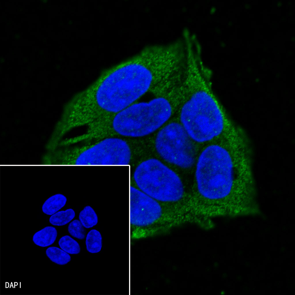
ICC shows positive staining in HepG2 cells. Anti-IGFBP1 antibody was used at 1/250 dilution (Green) and incubated overnight at 4°C. Goat polyclonal Antibody to Mouse IgG - H&L (Alexa Fluor® 488) was used as secondary antibody at 1/1000 dilution. The cells were fixed with 100% ice-cold methanol and permeabilized with 0.1% PBS-Triton X-100. Nuclei were counterstained with DAPI (Blue).
 您现在的位置: