

12 months from date of receipt / reconstitution, -20 °C as supplied
| 应用 | 稀释度 |
|---|---|
| WB | 1:1000 |
| IHC-P | 1:250 |
| ICFCM | 1:500 |
| ICC | 1:500 |
The androgen receptor (AR), ligand-induced transcription factor, is expressed in primary prostate cancer and in metastases. AR regulates multiple cellular events, proliferation, apoptosis, migration, invasion, and differentiation. Its expression in prostate cancer cells is regulated by steroid and peptide hormones [PMID: 24384911]. The elucidation of the structures of the AR DNA binding domain (DBD) and ligand binding domain (LBD) provides a new framework for understanding the functions of this receptor and leads to the development of rational drug design for the treatment of prostate cancer [PMID: 24909511].
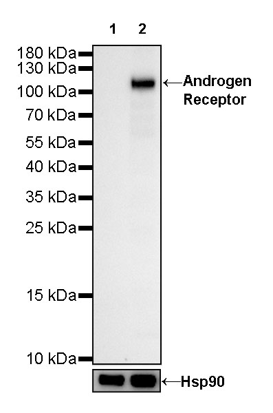
WB result of Androgen Receptor Rabbit mAb
Primary antibody: Androgen Receptor Rabbit mAb at 1/1000 dilution
Lane 1: PC-3 whole cell lysate 20 µg
Lane 2: LNCaP whole cell hot lysate with 1% SDS 20 µg
Negative control: PC-3 whole cell lysate
Secondary antibody: Goat Anti-Rabbit IgG, (H+L), HRP conjugated at 1/10000 dilution
Predicted MW: 98kDa
Observed MW: 120kDa

Flow cytometric analysis of 4% PFA fixed 90% methanol permeabilized LNCAP (H uman prostate carcinoma epithelial cell) cells labelling Androgen Receptor antibody at 1/500 (0.1 μg) dilution / (red) compared with a Rabbit monoclonal IgG (Black) isotype control and an unlabelled control (cells without incubation with primary antibody and secondary antibody) (Blue). Goat Anti - Rabbit IgG Alexa Fluor® 488 was used as the secondary antibody.

IHC shows positive staining in paraffin-embedded human testis. Anti-Androgen Receptor antibody was used at 1/250 dilution, followed by a HRP Polymer for Mouse & Rabbit IgG (ready to use). Counterstained with hematoxylin. Heat mediated antigen retrieval with Tris/EDTA buffer pH9.0 was performed before commencing with IHC staining protocol.

IHC shows positive staining in paraffin-embedded human breast cancer. Anti-Androgen Receptor antibody was used at 1/250 dilution, followed by a HRP Polymer for Mouse & Rabbit IgG (ready to use). Counterstained with hematoxylin. Heat mediated antigen retrieval with Tris/EDTA buffer pH9.0 was performed before commencing with IHC staining protocol.

IHC shows positive staining in paraffin-embedded human prostatic cancer. Anti-Androgen Receptor antibody was used at 1/250 dilution, followed by a HRP Polymer for Mouse & Rabbit IgG (ready to use). Counterstained with hematoxylin. Heat mediated antigen retrieval with Tris/EDTA buffer pH9.0 was performed before commencing with IHC staining protocol.
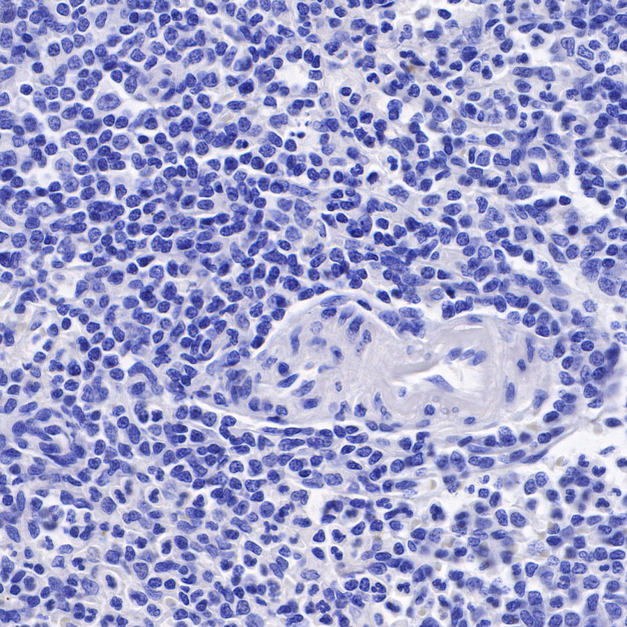
Negative control: IHC shows negative staining in paraffin-embedded human spleen. Anti-Androgen Receptor antibody was used at 1/250 dilution, followed by a HRP Polymer for Mouse & Rabbit IgG (ready to use). Counterstained with hematoxylin. Heat mediated antigen retrieval with Tris/EDTA buffer pH9.0 was performed before commencing with IHC staining protocol.

Negative control: IHC shows negative staining in paraffin-embedded human cervical squamous cell carcinoma. Anti-Androgen Receptor antibody was used at 1/250 dilution, followed by a HRP Polymer for Mouse & Rabbit IgG (ready to use). Counterstained with hematoxylin. Heat mediated antigen retrieval with Tris/EDTA buffer pH9.0 was performed before commencing with IHC staining protocol.
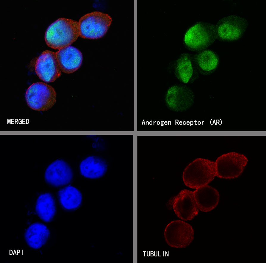
ICC shows positive staining in LnCap cells. Anti-Androgen Receptor antibody was used at 1/500 dilution (Green) and incubated overnight at 4°C. Goat polyclonal Antibody to Rabbit IgG - H&L (Alexa Fluor® 488) was used as secondary antibody at 1/1000 dilution. The cells were fixed with 100% ice-cold methanol and permeabilized with 0.1% PBS-Triton X-100. Nuclei were counterstained with DAPI (Blue).Counterstain with tubulin (red).
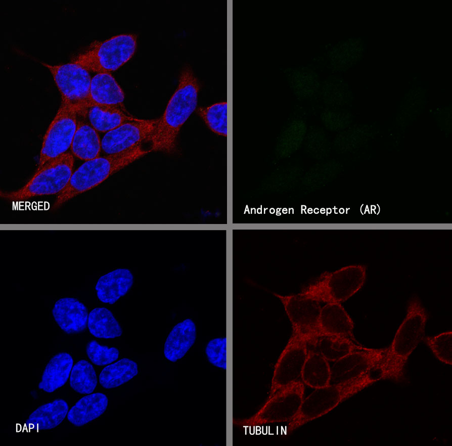
Negative control:ICC shows negative staining in 22Rv1 cells. Anti-Androgen Receptor antibody was used at 1/500 dilution and incubated overnight at 4°C. Goat polyclonal Antibody to Rabbit IgG - H&L (Alexa Fluor® 488) was used as secondary antibody at 1/1000 dilution. The cells were fixed with 100% ice-cold methanol and permeabilized with 0.1% PBS-Triton X-100. Nuclei were counterstained with DAPI (Blue).Counterstain with tubulin (red).
 您现在的位置: