

PBS pH7.4, containing no preservative
2 to 8 °C for 2 weeks under sterile conditions;
-20 °C for 3 months under sterile conditions;
-80 °C for 24 months under sterile conditions.
Please avoid repeated freeze-thaw cycles.
| 应用 | 稀释度 |
|---|---|
| WB | 1:250 |
| SPR | 2000nM-31.25nM |
| ICC | 1:500 |
Programmed cell death protein 1, also known as PD-1 and CD279 (cluster of differentiation 279), is a protein on the surface of T and B cells that has a role in regulating the immune system's response to the cells of the human body by down-regulating the immune system and promoting self-tolerance by suppressing T cell inflammatory activity. This prevents autoimmune diseases, but it can also prevent the immune system from killing cancer cells. PD-1 is an immune checkpoint and guards against autoimmunity through two mechanisms. First, it promotes apoptosis (programmed cell death) of antigen-specific T-cells in lymph nodes. Second, it reduces apoptosis in regulatory T cells (anti-inflammatory, suppressive T cells).
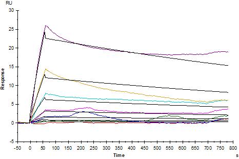
Anti-His antibody Immobilized on CM5 Chip captured PD1 His Tag, Mouse, can bind PD-1 Recombinant Rat mAb ( RMP1-14 ) with an affinity constant of 0.238 μM as determined in SPR assay.

WB result of PD-1 Rat mAb
Primary antibody: PD-1 Rat mAb at 1/250 dilution
Lane 1:PD-1 Fc Chimera, Mouse (recombinant protein) lysate 1 μg
Secondary antibody: Goat Anti-Rat IgG, (H+L), HRP conjugated at 1/10000 dilution
Predicted MW: 58~70 kDa
Observed MW: 58~70 kDa
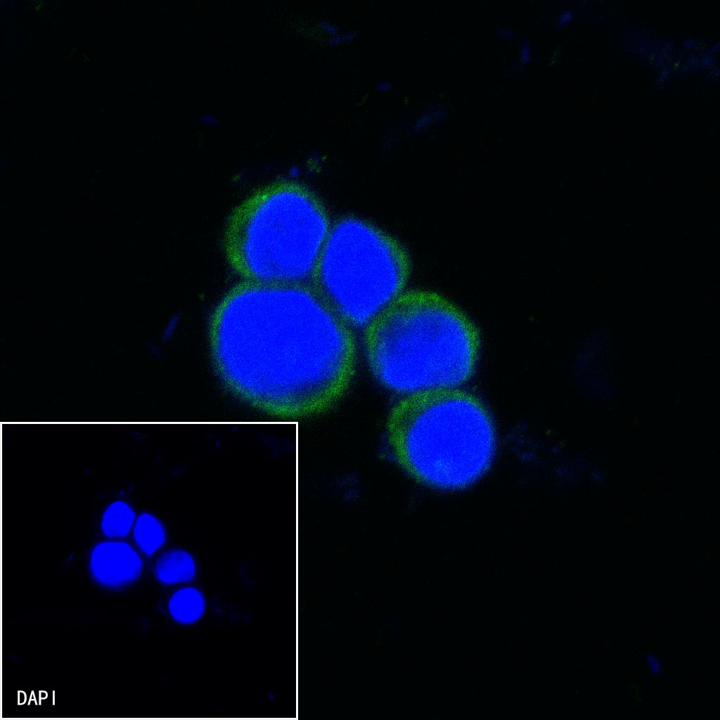
ICC shows positive staining in EL4.IL2 cells. Anti-PD-1 antibody was used at 1/500 dilution (Green) and incubated overnight at 4°C. Goat polyclonal Antibody to rat IgG - H&L (Alexa Fluor® 488) was used as secondary antibody at 1/1000 dilution. The cells were fixed with 100% ice-cold methanol and permeabilized with 0.1% PBS-Triton X-100. Nuclei were counterstained with DAPI (Blue).
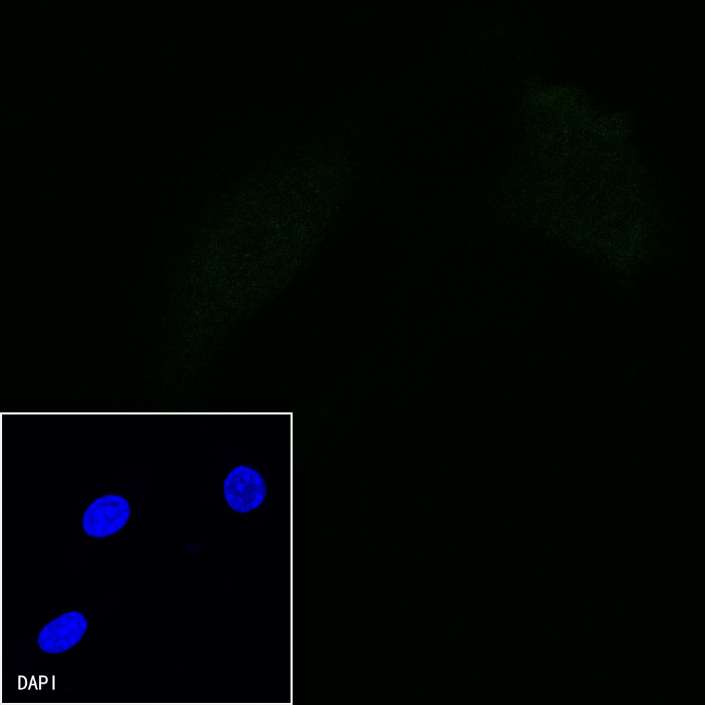
Negative control:ICC shows negative staining in NIH/3T3 cells. Anti-PD-1 antibody was used at 1/500 dilution and incubated overnight at 4°C. Goat polyclonal Antibody to rat IgG - H&L (Alexa Fluor® 488) was used as secondary antibody at 1/1000 dilution. The cells were fixed with 100% ice-cold methanol and permeabilized with 0.1% PBS-Triton X-100. Nuclei were counterstained with DAPI (Blue).
 您现在的位置: