

| 应用 | 稀释度 |
|---|---|
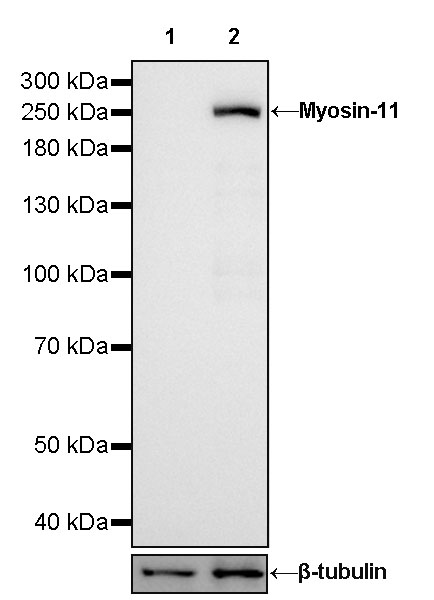
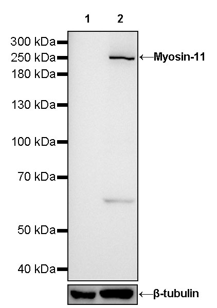
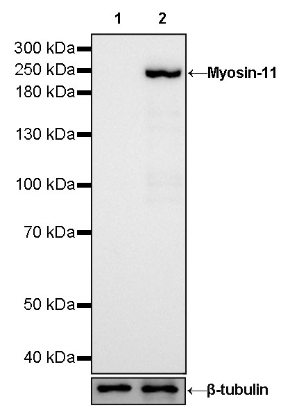
| WB | 1:1000-1:25000 |
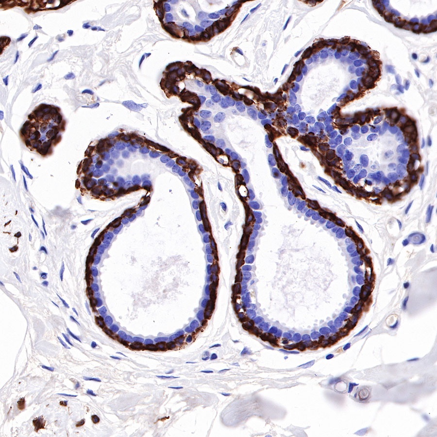
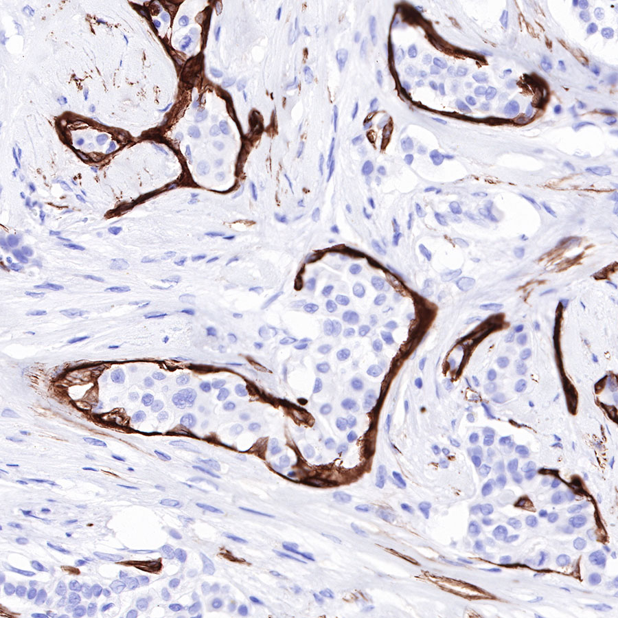
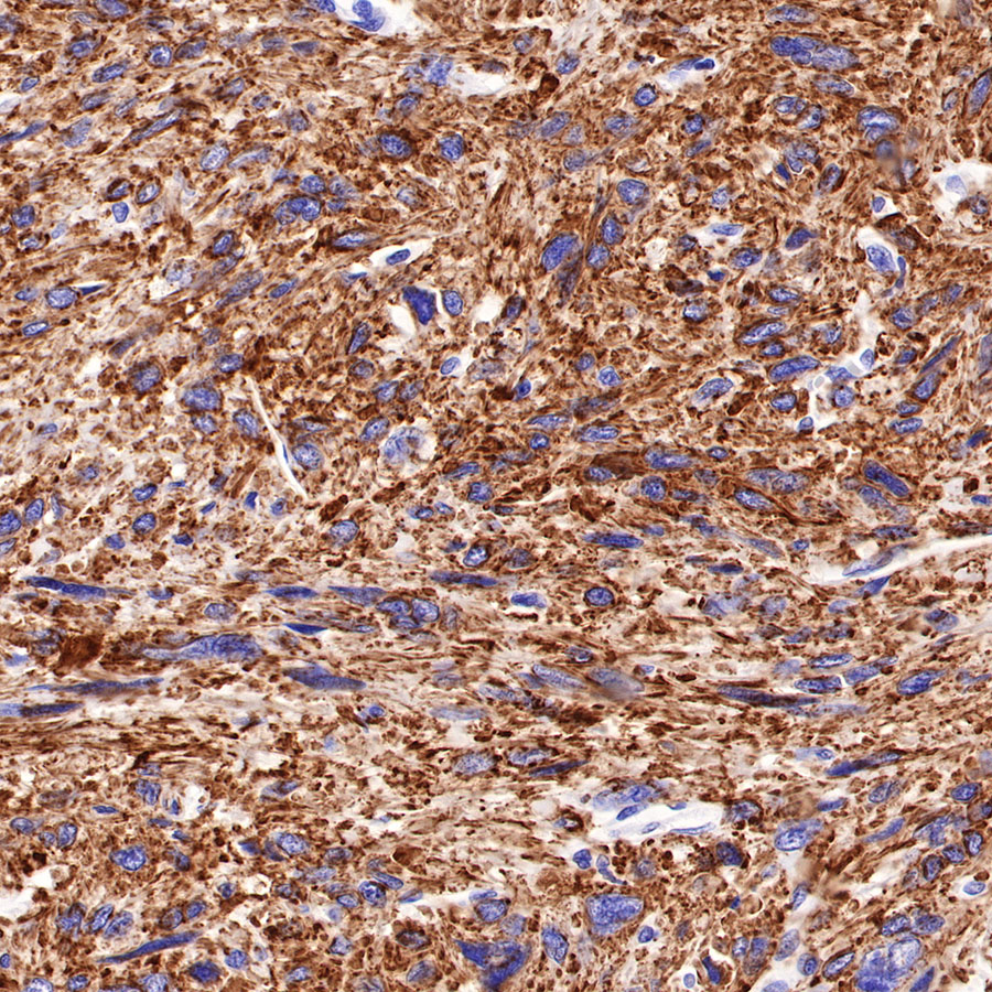
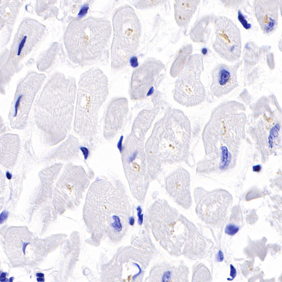
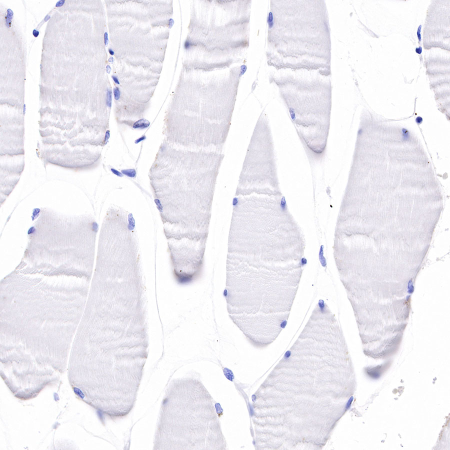
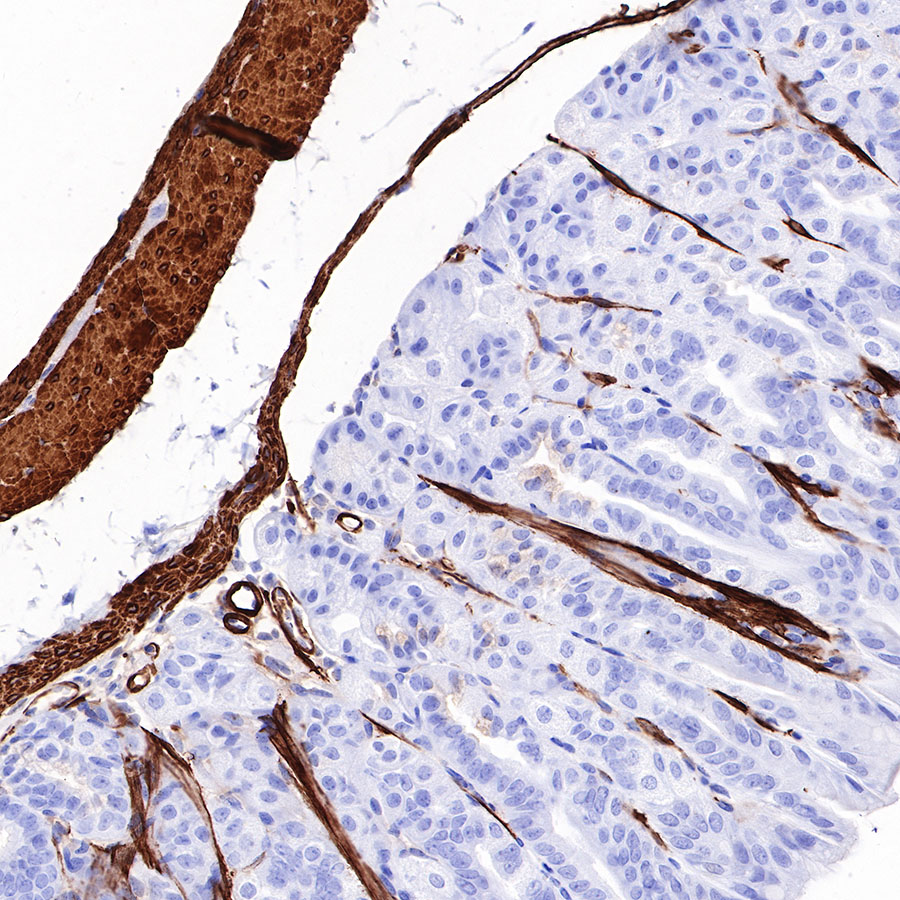
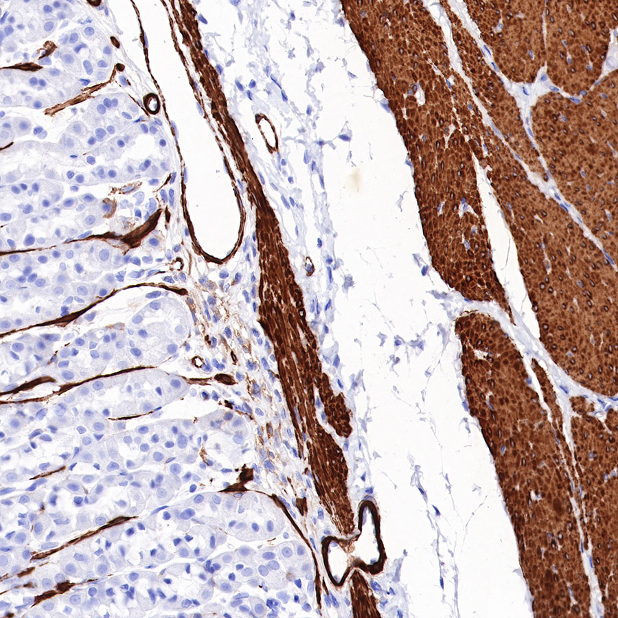
| IHC-P | 1:1000 |
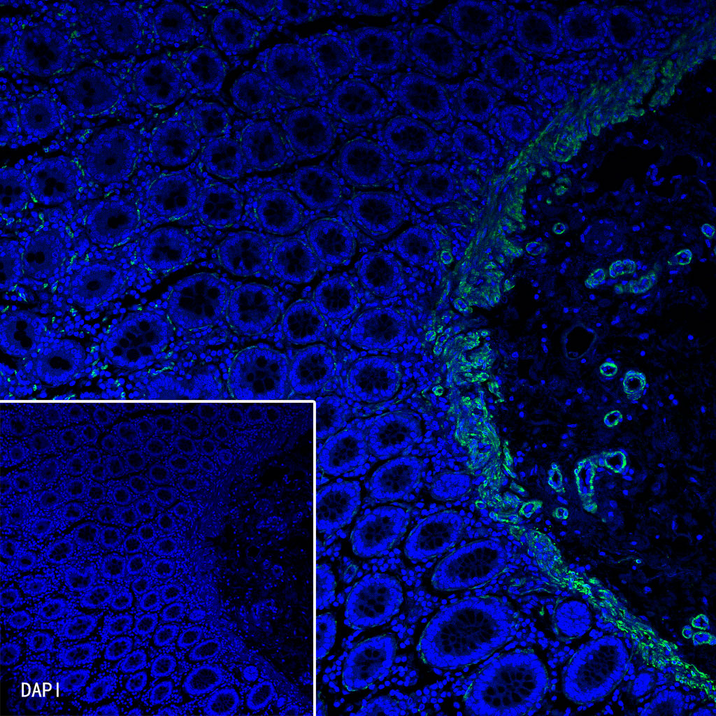
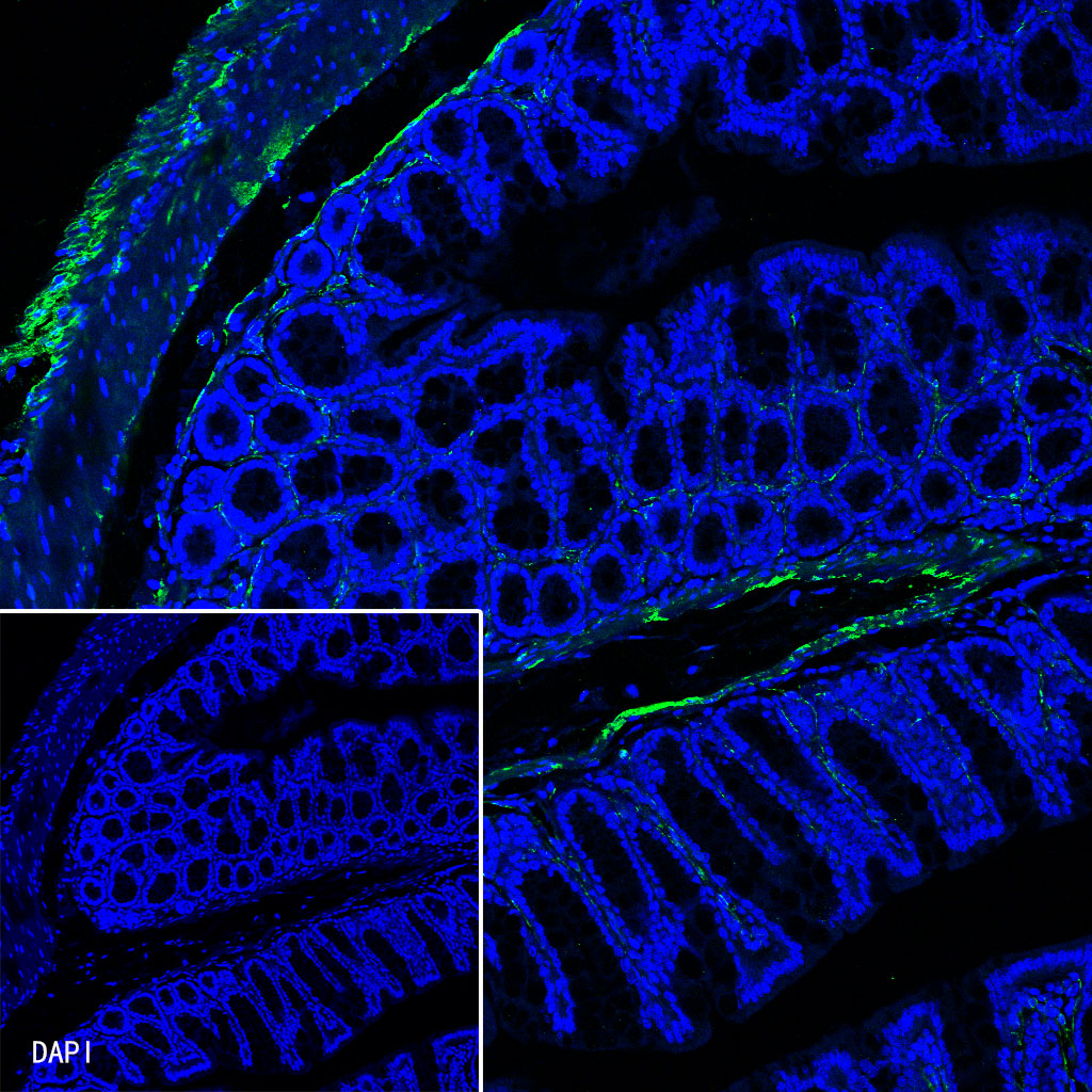
| IF | 1:125 |
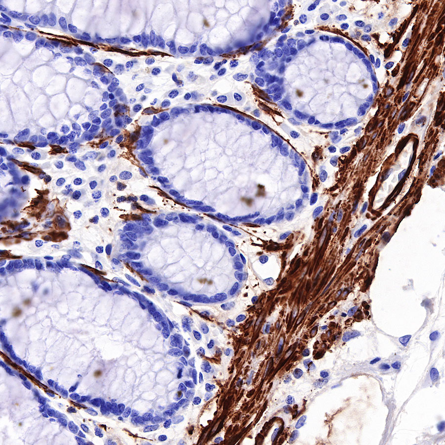
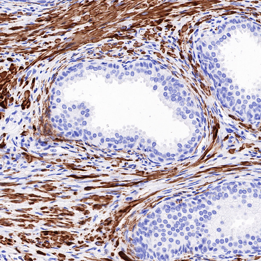
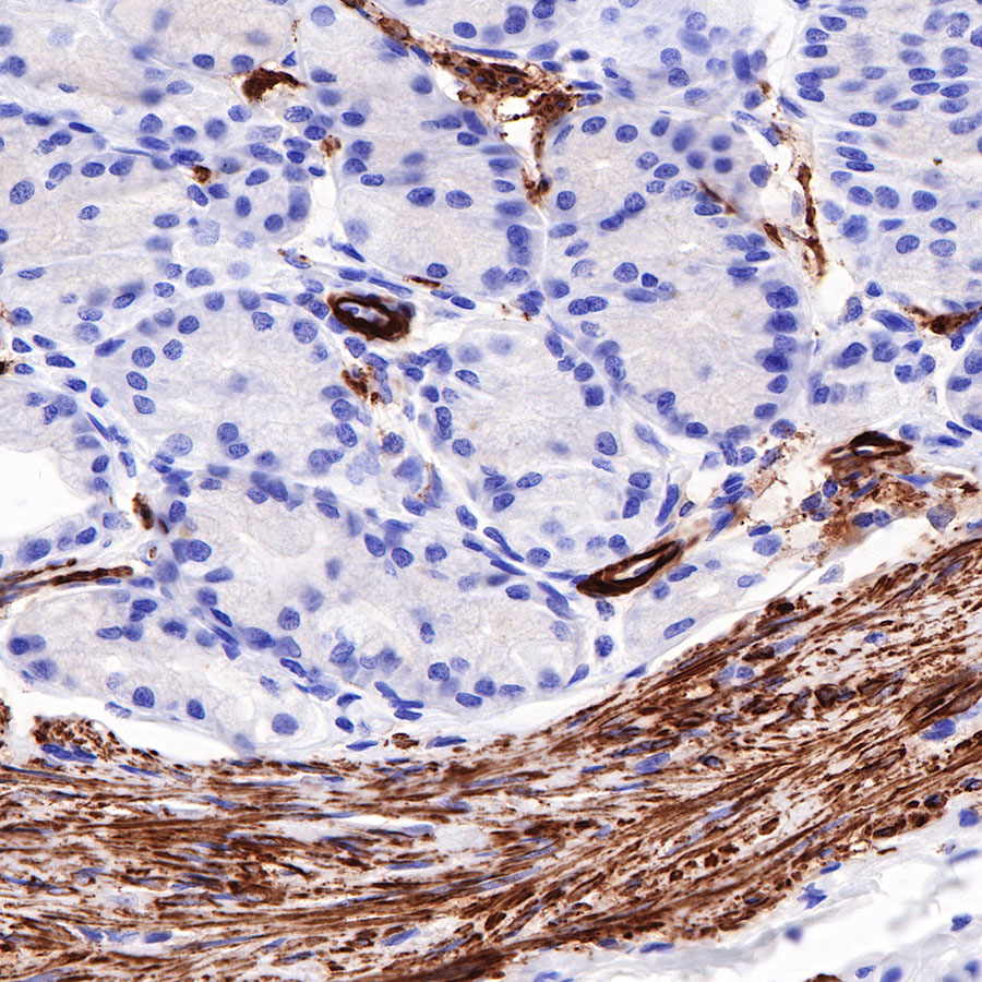
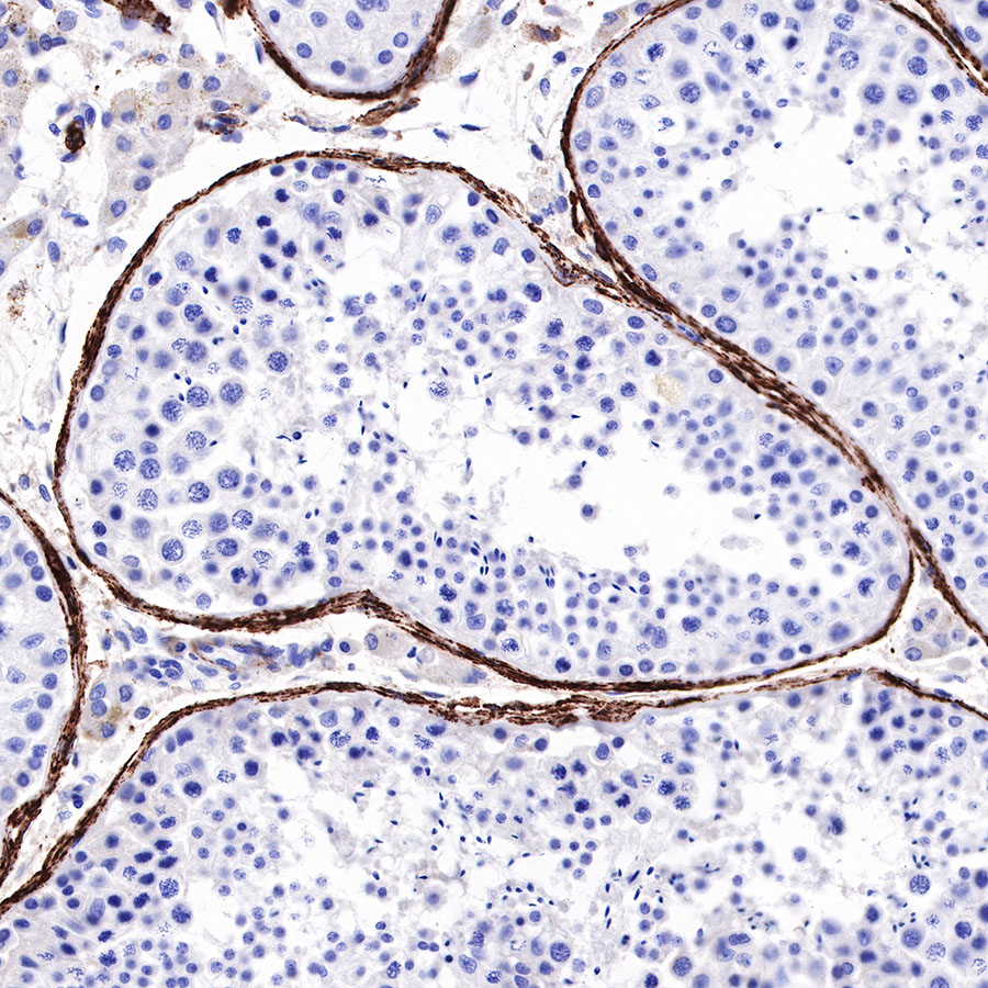
SMMHC is a structural component of the smooth muscle myosin molecule and is a specific marker of “terminal” smooth muscle differentiation [PMID: 10721417, PMID: 3882826]. SMMHC is composed of at least two isoforms: SM1 (204 kDa) and SM2 (200 kDa), both of which are encoded by a single gene [PMID: 9702855, PMID: 10998642]. The SM1 isoform is expressed in the MEC of normal mammary glands, fibrocystic diseases, and in myoepithelial derived tumours of the breast [PMID: 11493962, PMID: 9702855]. Furthermore, studies have documented that antibodies to SMMHC and calponin, both markers of terminal smooth muscle differentiation, are more specific for breast MEC than are other more commonly used antibodies, such as those that recognise SMA [PMID: 11493962].
 您现在的位置: