
PBS, 40% Glycerol, 0.05% BSA, 0.03% Proclin 300
12 months from date of receipt / reconstitution, -20 °C as supplied
| 应用 | 稀释度 |
|---|---|
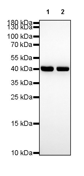
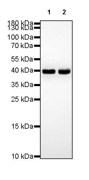
| WB | 1:1000-1:5000 |
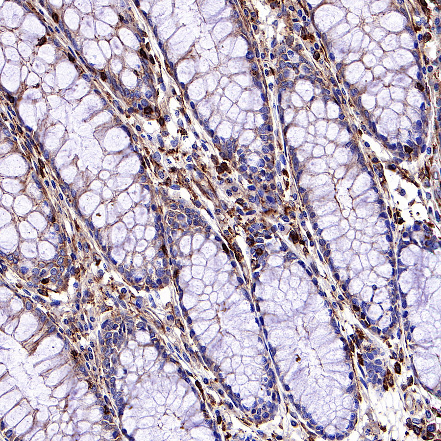
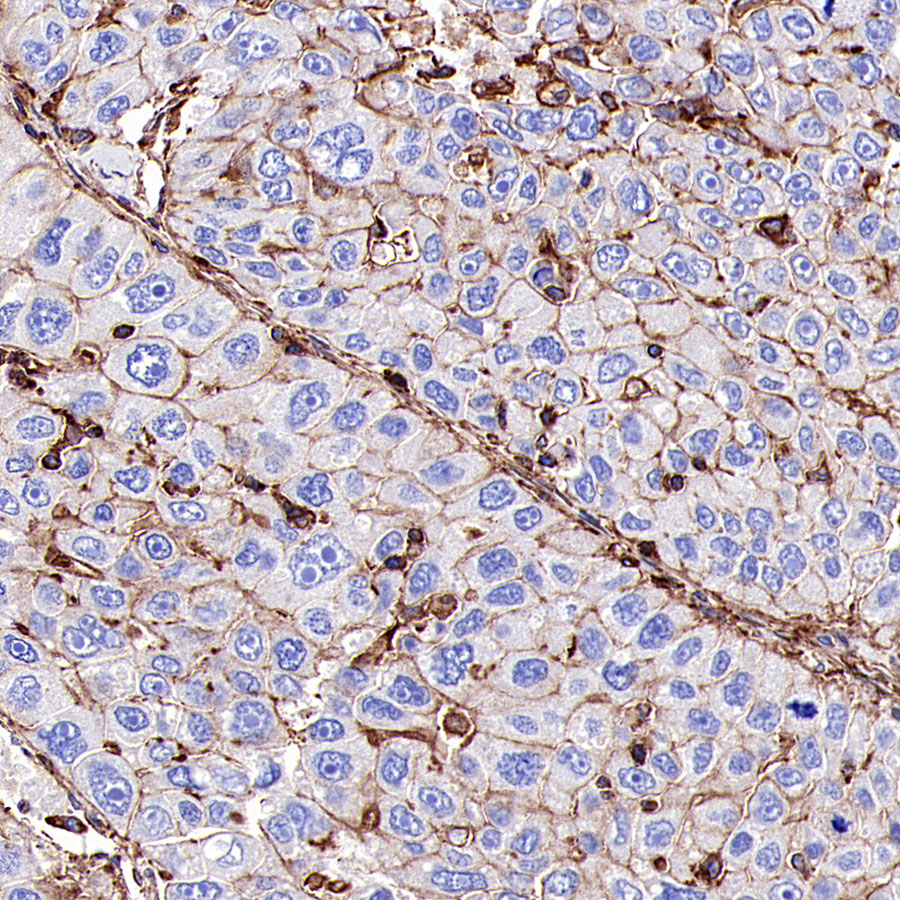
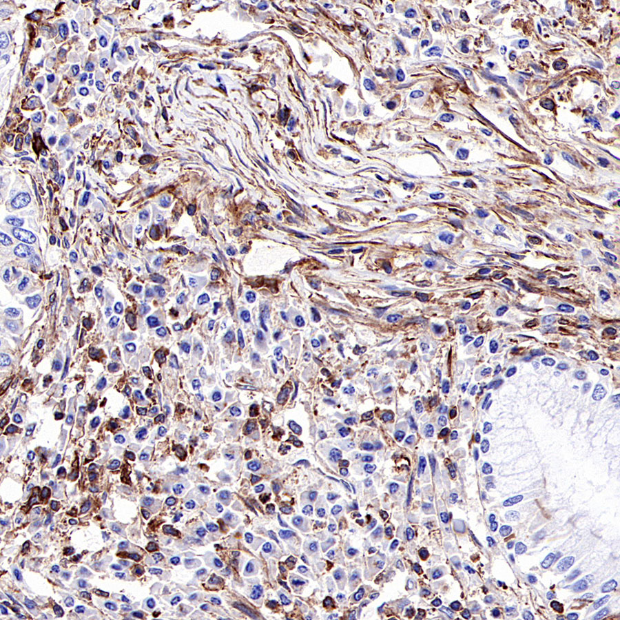
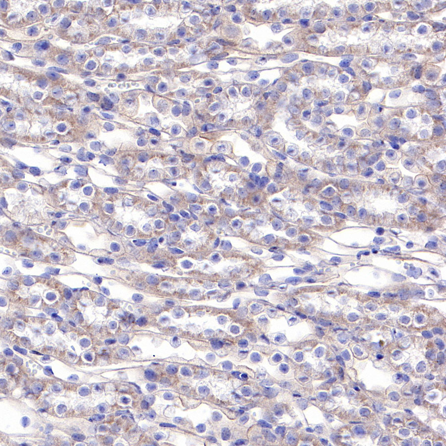
| IHC-P | 1:2000 |
| ICC | 1:500 |
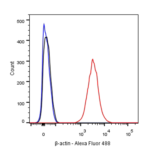
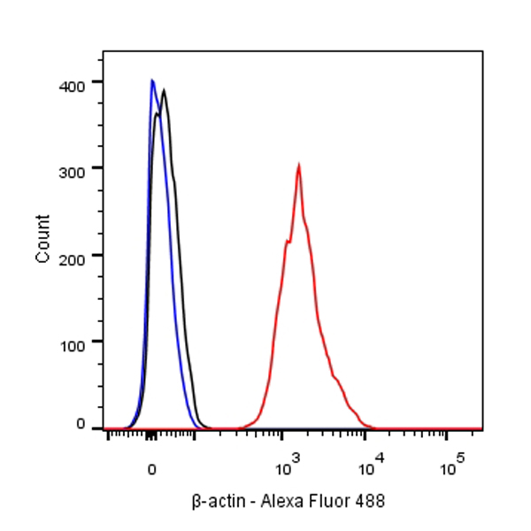
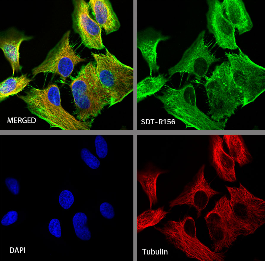
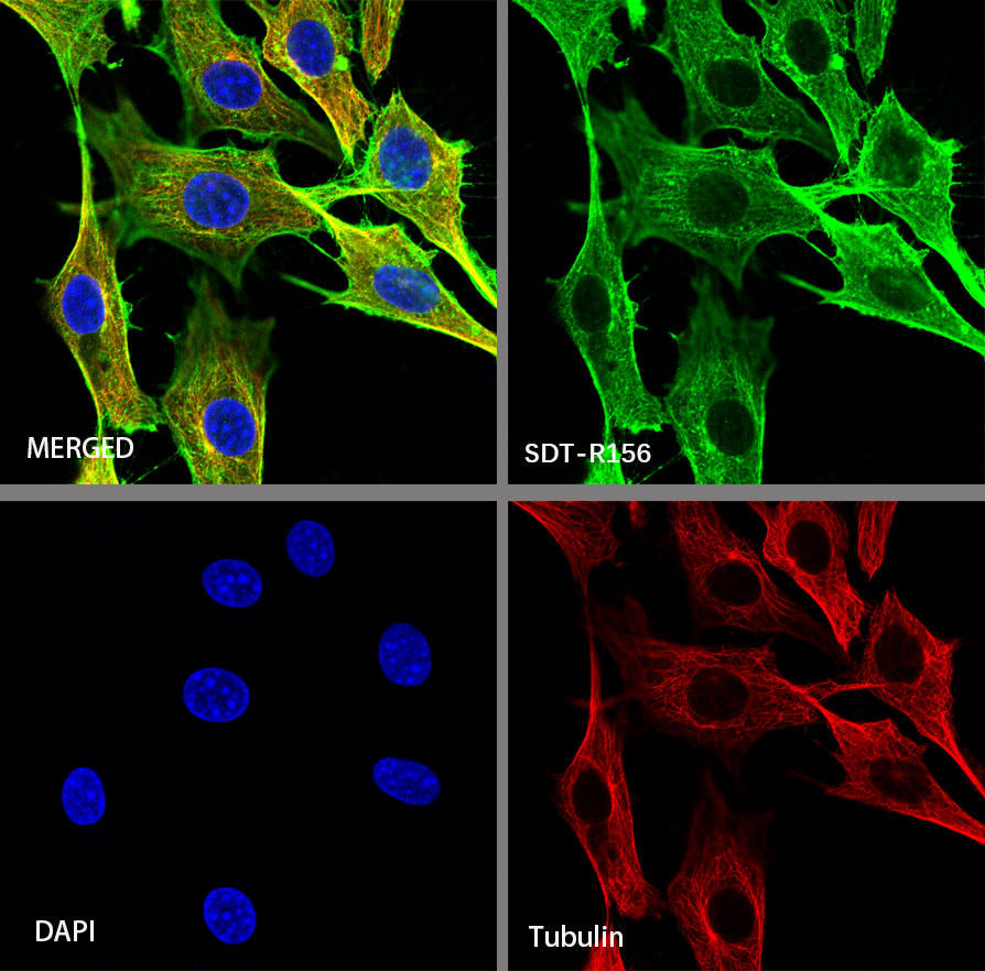
| ICFCM | 1:500 |
Beta-actin (human gene and protein abbreviation ACTB/ACTB) is one of six different actin isoforms which have been identified in humans. This is one of the two non-muscle cytoskeletal actins. Actins are highly conserved proteins that are involved in cell motility, structure and integrity. Beta actin is often used in Western blotting as a loading control, to normalize total protein amounts and check for eventual protein degradation in the samples. Its transcript is also commonly used as a housekeeping gene standard in qPCR. Its molecular weight is approximately 42 kDa.


 您现在的位置: