
12 months from date of receipt / reconstitution, -20 °C as supplied
| 应用 | 稀释度 |
|---|---|
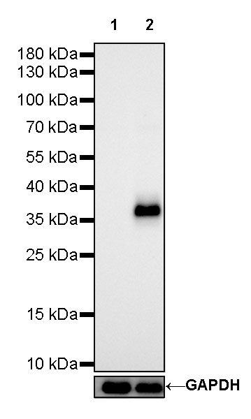
| WB | 1:500 |
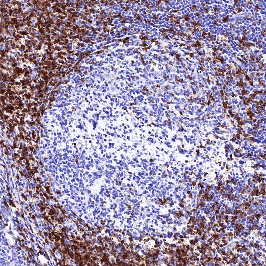
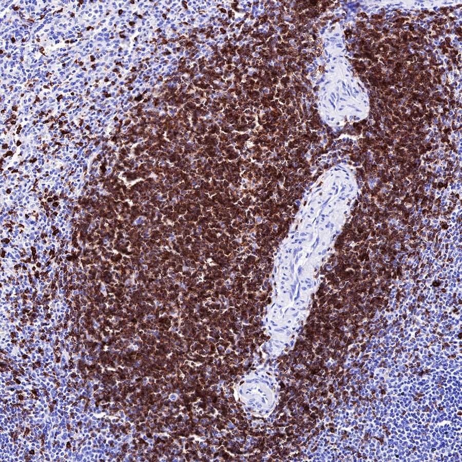
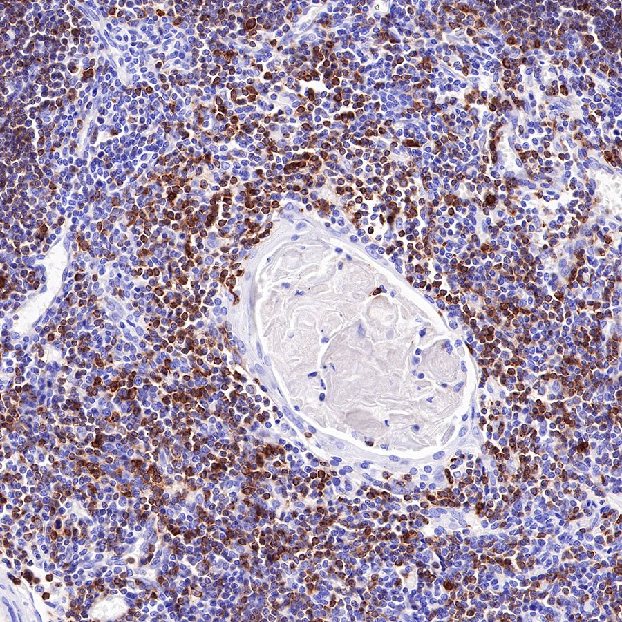
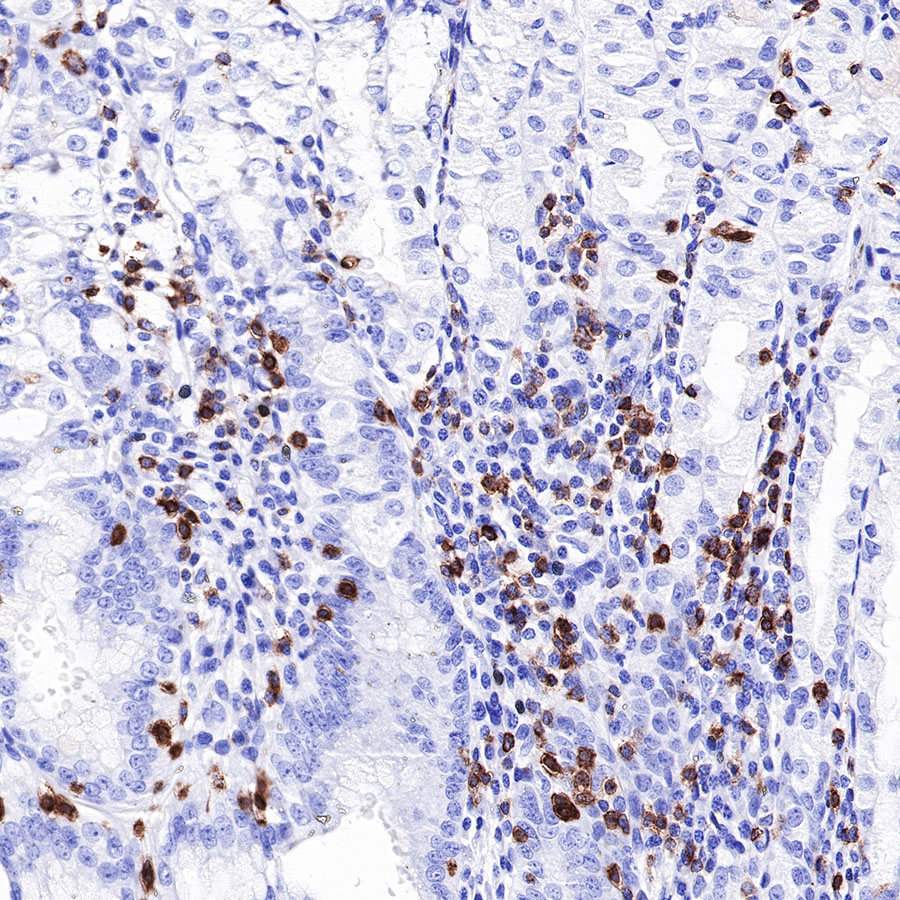
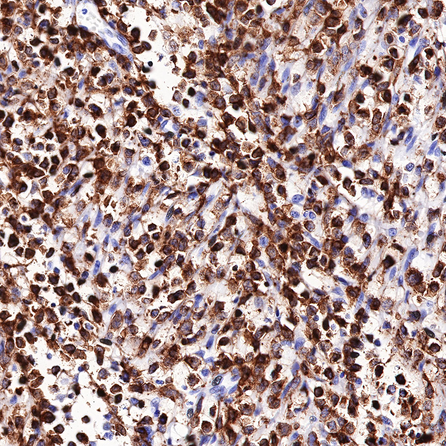
| IHC-P | 1:1000 |
| ICC | 1:125 |
| IP | 1:25 |
CD7 is a single-domain Ig superfamily molecule expressed on human T and NK cells, as well as on cells in the early stages of T, B, and myeloid cell differentiation. CD7 is highly expressed on malignant immature T cells and is generally absent on malignant mature T cells [PMID: 10530432]. CD7 is Lymphoid marker, which expressed in 30% of AML cases and linked with poor prognosis in myeloid malignancies [PMID: 21148082].

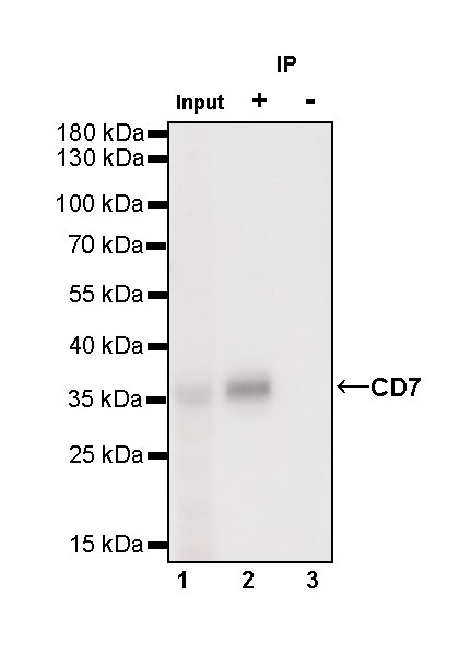
CD7 Rabbit mAb at 1/25 dilution (1 µg) immunoprecipitating CD7 in 0.4 mg Jurkat whole cell lysate.
Western blot was performed on the immunoprecipitate using CD7 Rabbit mAb at 1/1000 dilution.
Secondary antibody (HRP) for IP was used at 1/400 dilution.
Lane 1: Jurkat whole cell lysate 50 µg (Input)
Lane 2: CD7 Rabbit mAb IP in Jurkat whole cell lysate
Lane 3: Rabbit monoclonal IgG IP in Jurkat whole cell lysate
Predicted MW: 25.4 kDa
Observed MW: 37 kDa


 您现在的位置: