
PBS, 40% Glycerol, 0.05% BSA, 0.03% Proclin 300
12 months from date of receipt / reconstitution, -20 °C as supplied
| 应用 | 稀释度 |
|---|---|
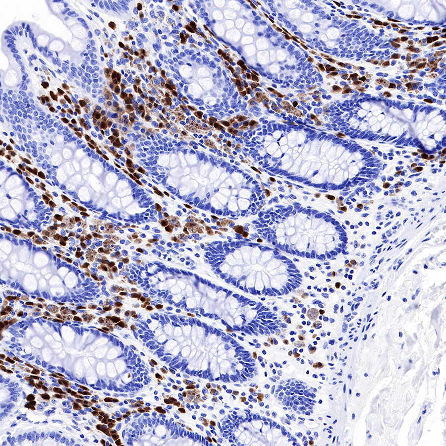
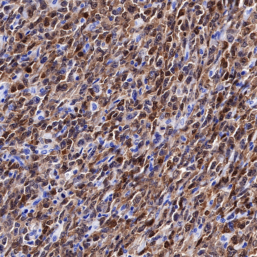
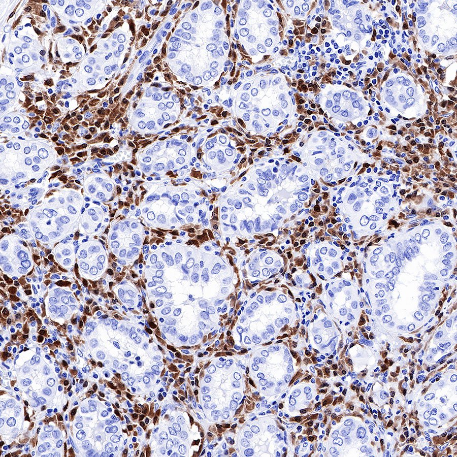
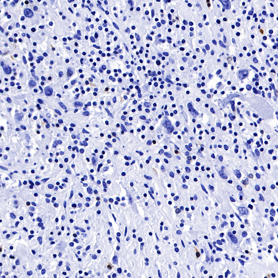
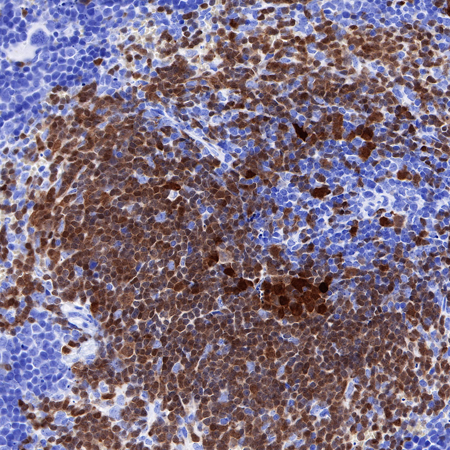
| IHC-P | 1:500 |
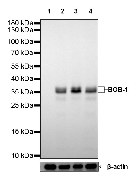
| WB | 1:5000 |
| ICC | 1:125 |
BOB-1 interacts with the sequence-specific DNA-binding POU transcription factors (named after the founding family members PIT1, OCT1/2, and UNC86), the ubiquitously expressed OCT1 (POU2F1) and lymphoid-specific OCT2 (POU2F2). As a transcriptional co-activator, BOB-1 itself does not bind DNA but is rather recruited into transcriptional regulation via interaction with DNA-bound POU-domain transcription factors OCT1 and OCT2. The POU-domain is a unique bipartite structure allowing DNA recognition with remarkable flexibility .


ICC shows positive staining in Ramos cells. Anti-BOB-1 antibody was used at 1/125 dilution (Green) and incubated overnight at 4°C. Goat polyclonal Antibody to Rabbit IgG - H&L (Alexa Fluor® 488) was used as secondary antibody at 1/1000 dilution. The cells were fixed with 4% PFA and permeabilized with 0.1% PBS-Triton X-100. Nuclei were counterstained with DAPI (Blue).
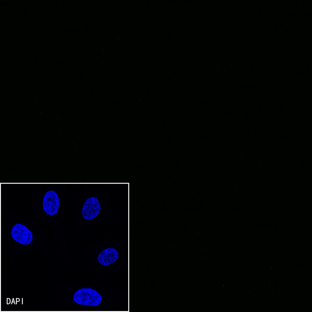
Negative control:ICC shows negative staining in HeLa cells. Anti-BOB-1 antibody was used at 1/125 dilution and incubated overnight at 4°C. Goat polyclonal Antibody to Rabbit IgG - H&L (Alexa Fluor® 488) was used as secondary antibody at 1/1000 dilution. The cells were fixed with 4% PFA and permeabilized with 0.1% PBS-Triton X-100. Nuclei were counterstained with DAPI (Blue).
 您现在的位置: