
12 months from date of receipt / reconstitution, -20 °C as supplied
| 应用 | 稀释度 |
|---|---|
| WB | 1:1000 |
| IHC-P | 1:500 |
| ICFCM | 1:500 |
| ICC | 1:500 |
Progesterone receptor (PR) is a master regulator in female reproductive tissues that controls developmental processes and proliferation and differentiation during the reproductive cycle and pregnancy. PR also plays a role in progression of endocrine-dependent breast cancer. As a member of the nuclear receptor family of ligand-dependent transcription factors, the main action of PR is to regulate networks of target gene expression in response to binding its cognate steroid hormone, progesterone [PMID: 27380738].

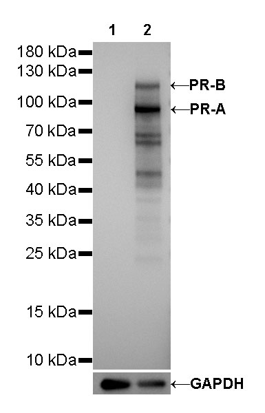
WB result of Progesterone Receptor Rabbit mAb
Primary antibody: Progesterone Receptor Rabbit mAb at 1/1000 dilution
Lane 1: MDA-MB-231 whole cell lysate 20 µg
Lane 2: T-47D whole cell lysate 20 µg
Negative control: MDA-MB-231 whole cell lysate
Secondary antibody: Goat Anti-Rabbit IgG, (H+L), HRP conjugated at 1/10000 dilution
Predicted MW: 82, 99 kDa
Observed MW: 90, 118 kDa
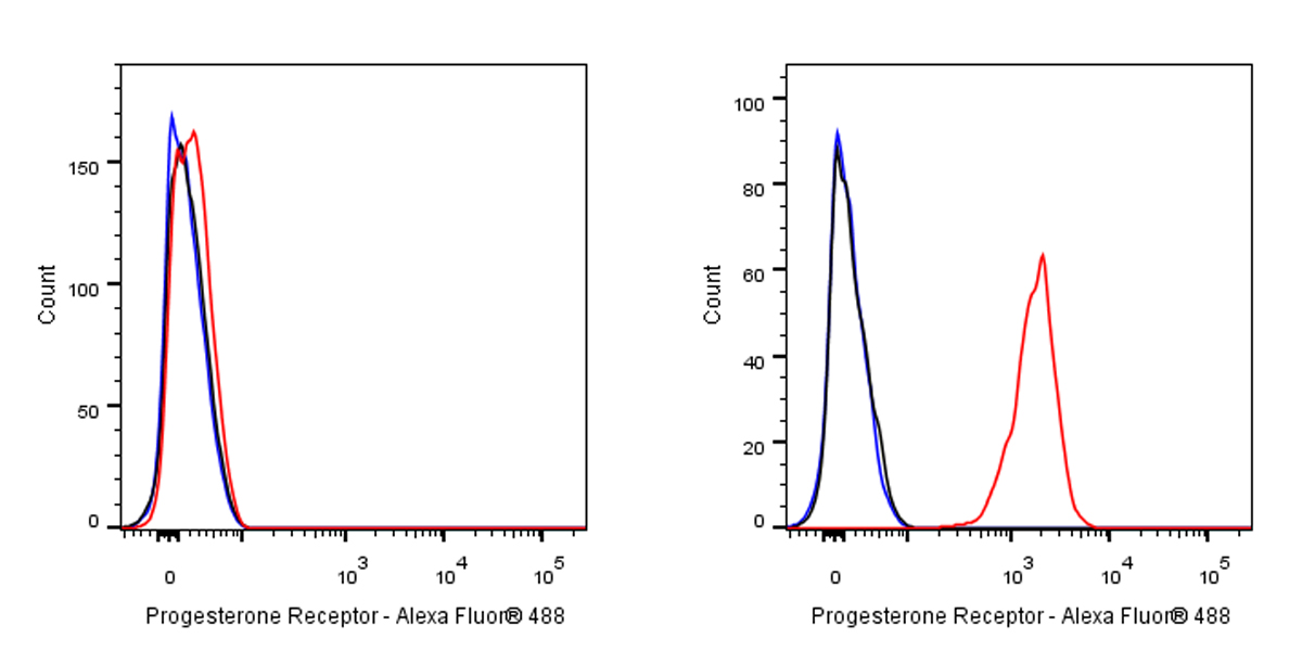
Flow cytometric analysis of 4% PFA fixed 90% methanol permeabilized MDA-MB-231 (Human breast adenocarcinoma epithelial cell) / T47D (Human ductal breast epithelial tumor epithelial cell, right) cells labelling Androgen Receptor antibody at 1/500 dilution (0.1 μg) / (red) compared with a Rabbit monoclonal IgG (Black) isotype control and an unlabelled control (cells without incubation with primary antibody and secondary antibody) (Blue). Goat Anti - Rabbit IgG Alexa Fluor® 488 was used as the secondary antibody.
Negative control: MDA-MB-231
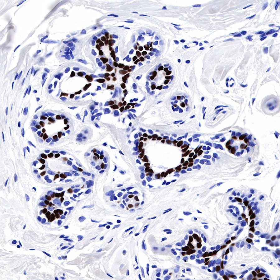
IHC shows positive staining in paraffin-embedded human breast. Anti-Progesterone Receptor antibody was used at 1/500 dilution, followed by a HRP Polymer for Mouse & Rabbit IgG (ready to use). Counterstained with hematoxylin. Heat mediated antigen retrieval with Tris/EDTA buffer pH9.0 was performed before commencing with IHC staining protocol.
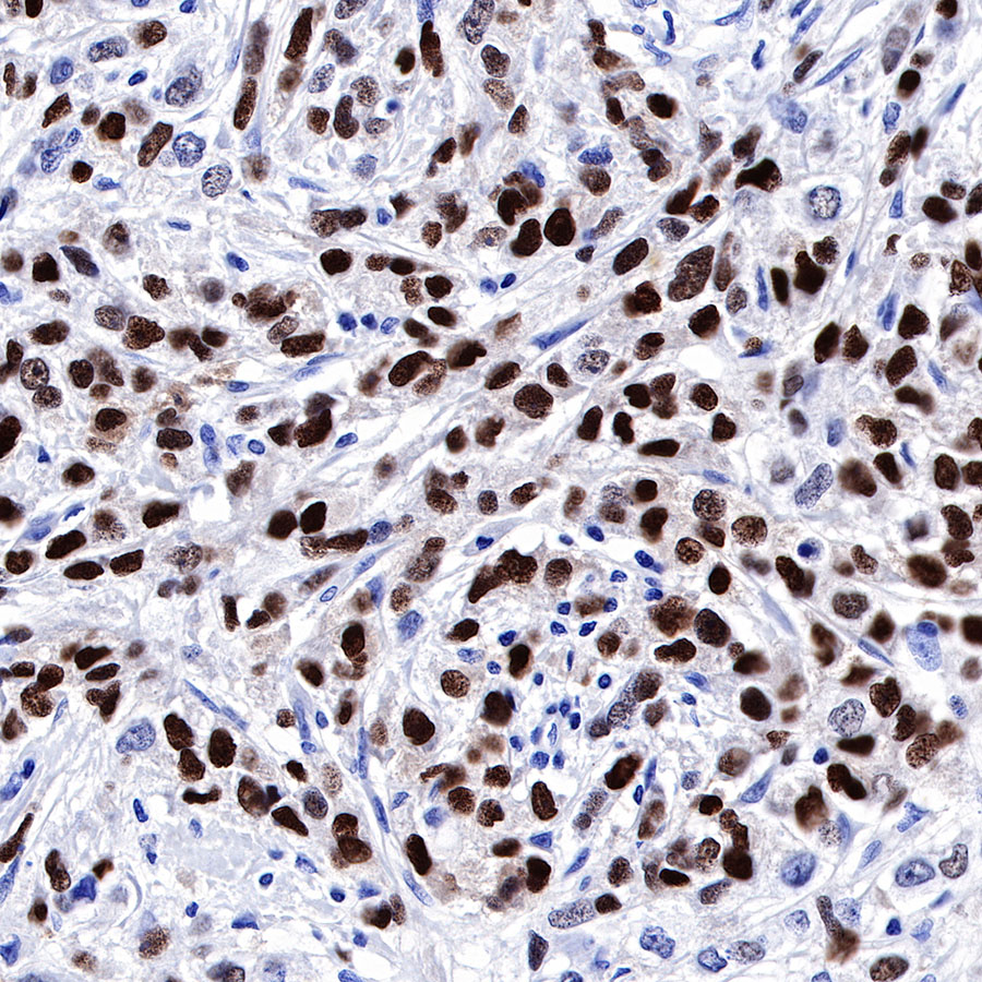
IHC shows positive staining in paraffin-embedded human breast cancer. Anti-Progesterone Receptor antibody was used at 1/500 dilution, followed by a HRP Polymer for Mouse & Rabbit IgG (ready to use). Counterstained with hematoxylin. Heat mediated antigen retrieval with Tris/EDTA buffer pH9.0 was performed before commencing with IHC staining protocol.
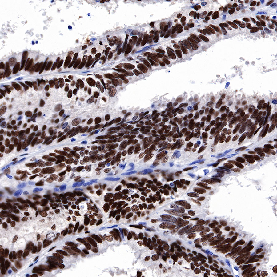
IHC shows positive staining in paraffin-embedded human endometrial cancer. Anti-Progesterone Receptor antibody was used at 1/500 dilution, followed by a HRP Polymer for Mouse & Rabbit IgG (ready to use). Counterstained with hematoxylin. Heat mediated antigen retrieval with Tris/EDTA buffer pH9.0 was performed before commencing with IHC staining protocol.
IHC shows positive staining in paraffin-embedded human ovarian cancer. Anti-Progesterone Receptor antibody was used at 1/500 dilution, followed by a HRP Polymer for Mouse & Rabbit IgG (ready to use). Counterstained with hematoxylin. Heat mediated antigen retrieval with Tris/EDTA buffer pH9.0 was performed before commencing with IHC staining protocol.
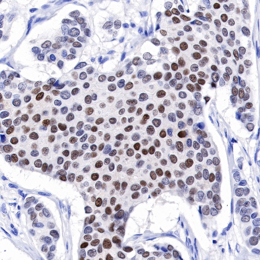
IHC shows positive staining in paraffin-embedded human breast cancer. Anti-Progesterone Receptor antibody was used at 1/500 dilution, followed by a HRP Polymer for Mouse & Rabbit IgG (ready to use). Counterstained with hematoxylin. Heat mediated antigen retrieval with Tris/EDTA buffer pH9.0 was performed before commencing with IHC staining protocol.

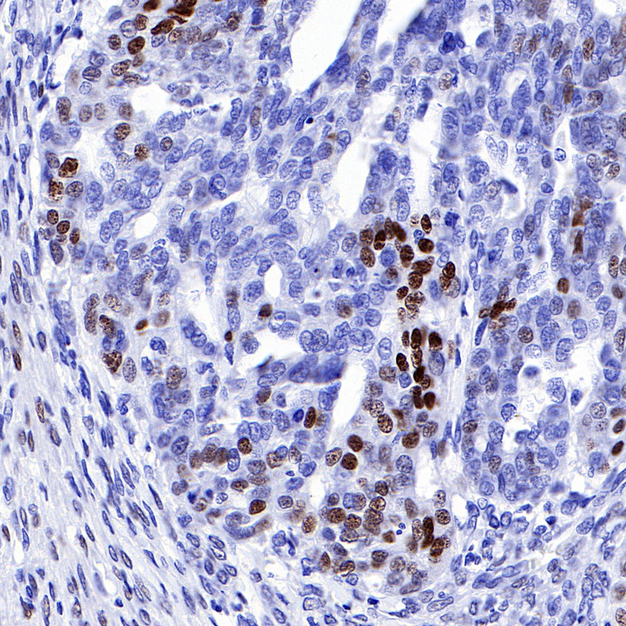
Negative control: IHC shows negative staining in paraffin-embedded human tonsil. Anti-Progesterone Receptor antibody was used at 1/500 dilution, followed by a HRP Polymer for Mouse & Rabbit IgG (ready to use). Counterstained with hematoxylin. Heat mediated antigen retrieval with Tris/EDTA buffer pH9.0 was performed before commencing with IHC staining protocol.
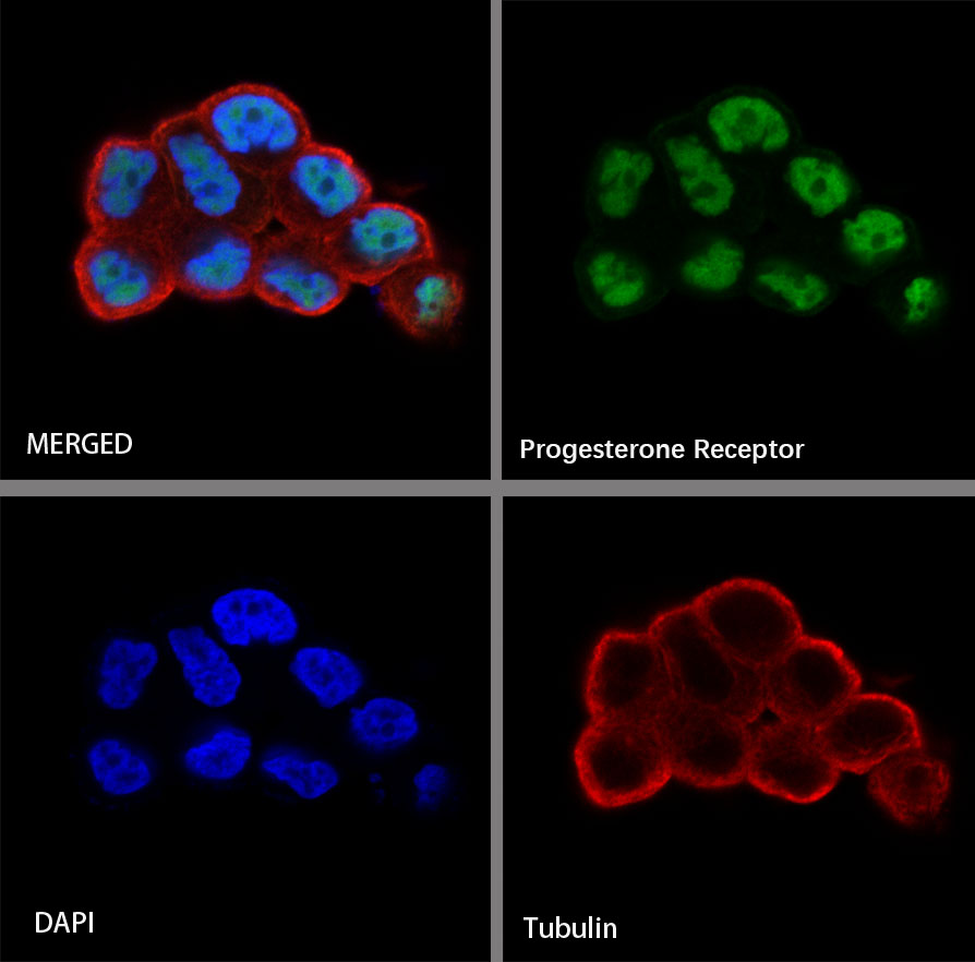
ICC shows positive staining in T47-D cells. Anti-Progesterone Receptor antibody was used at 1/500 dilution (Green) and incubated overnight at 4°C. Goat polyclonal Antibody to Rabbit IgG - H&L (Alexa Fluor® 488) was used as secondary antibody at 1/1000 dilution. The cells were fixed with 4% PFA and permeabilized with 0.1% PBS-Triton X-100. Nuclei were counterstained with DAPI (Blue). Counterstain with tubulin (red).
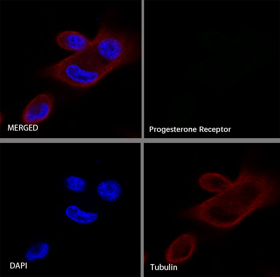
Negative control:ICC shows negative staining in MDA-MB-231 cells. Anti-Progesterone Receptor antibody was used at 1/500 dilution and incubated overnight at 4°C. Goat polyclonal Antibody to Rabbit IgG - H&L (Alexa Fluor® 488) was used as secondary antibody at 1/1000 dilution. The cells were fixed with 4% PFA and permeabilized with 0.1% PBS-Triton X-100. Nuclei were counterstained with DAPI (Blue). Counterstain with tubulin (red).
 您现在的位置: