

12 months from date of receipt / reconstitution, -20 °C as supplied
| 应用 | 稀释度 |
|---|---|
| WB | 1:5000 |
| IHC-P | 1:1000 |
| IP | 1:25 |
| ICFCM | 1:250 |
| ICC | 1:250 |
Thrombomodulin (TM) is an integral component of a multimolecular system, localized primarily to the vascular endothelium, that integrates crucial biological processes and biochemical pathways, including those related to coagulation, innate immunity, inflammation, and cell proliferation. These are designed to protect the host from injury and promote healing [PMID: 29866818].
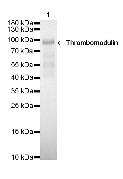
WB result of Thrombomodulin Rabbit mAb
Primary antibody: Thrombomodulin Rabbit mAb at 1/5000 dilution
Lane 1: A431 whole cell lysate 5 µg
Secondary antibody: Goat Anti-Rabbit IgG, (H+L), HRP conjugated at 1/10000 dilution
Predicted MW: 60 kDa
Observed MW: 90 kDa
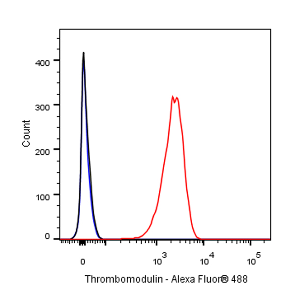
Flow cytometric analysis of A431 (Human epidermoid carcinoma epithelial cell) cells labelling Thrombomodulin antibody at 1/250 dilution (0.1 μg)/ (Red) compared with a Rabbit monoclonal IgG (Black) isotype control and an unlabelled control (cells without incubation with primary antibody and secondary antibody) (Blue). Goat Anti - Rabbit IgG Alexa Fluor® 488 was used as the secondary antibody.

Thrombomodulin Rabbit mAb at 1/25 dilution (1 µg) immunoprecipitating Thrombomodulin in 0.4 mg A431 whole cell lysate.
Western blot was performed on the immunoprecipitate using Thrombomodulin Rabbit mAb at 1/5000 dilution.
Secondary antibody (HRP) for IP was used at 1/400 dilution.
Lane 1: A431 whole cell ly-sate 10 µg (Input)
Lane 2: Thrombomodulin Rabbit mAb IP in A431 whole cell lysate
Lane 3: Rabbit monoclonal IgG IP in A431 whole cell lysate
Predicted MW: 60 kDa
Observed MW: 90 kDa
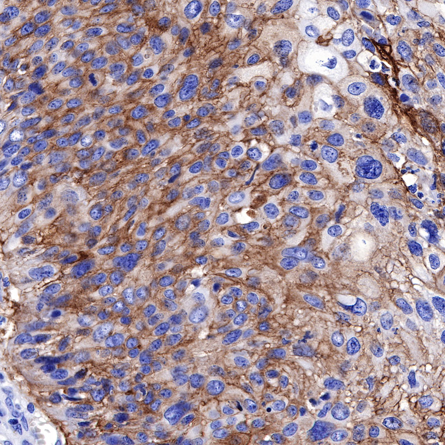
IHC shows positive staining in paraffin-embedded human cervcial squamous cell carcinoma. Anti-Thrombomodulin antibody was used at 1/1000 dilution, followed by a HRP Polymer for Mouse & Rabbit IgG (ready to use). Counterstained with hematoxylin. Heat mediated antigen retrieval with Tris/EDTA buffer pH9.0 was performed before commencing with IHC staining protocol.
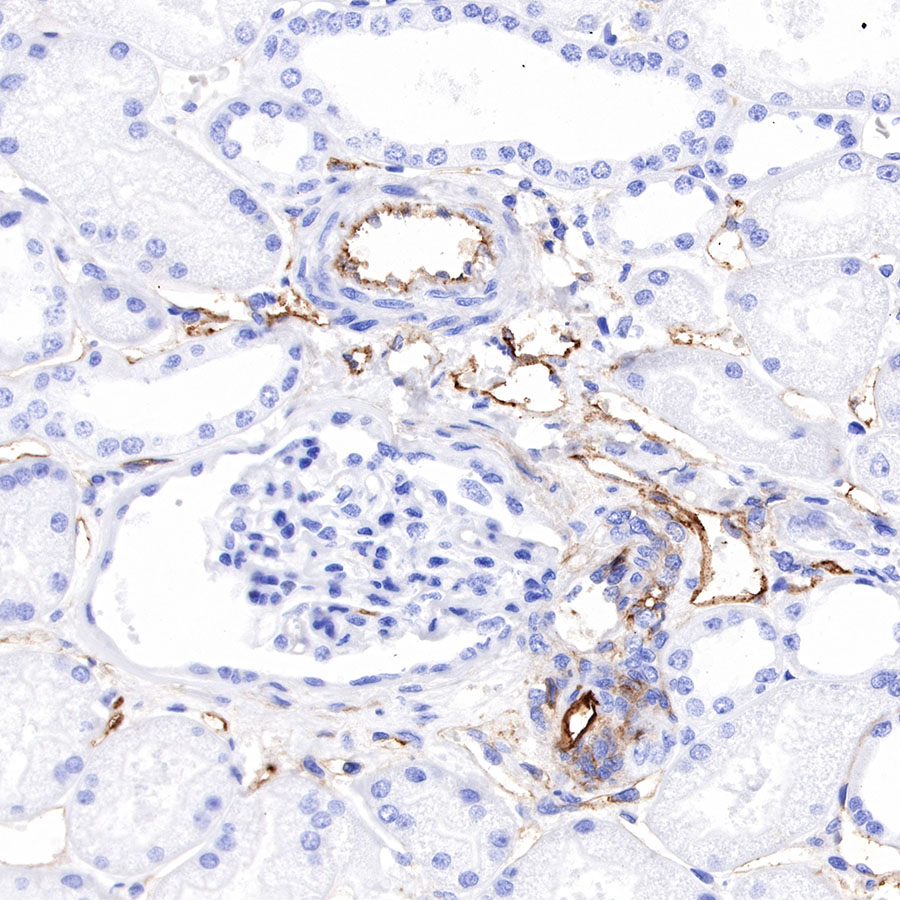
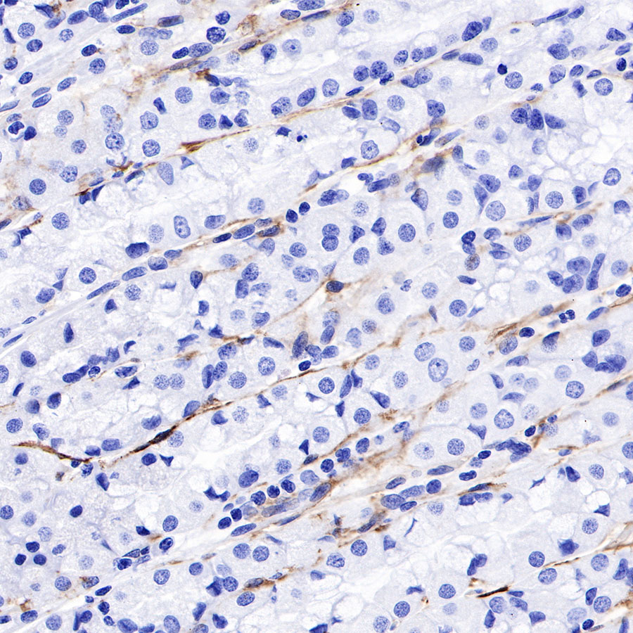
IHC shows positive staining in paraffin-embedded human kidney. Anti-Thrombomodulin antibody was used at 1/1000 dilution, followed by a HRP Polymer for Mouse & Rabbit IgG (ready to use). Counterstained with hematoxylin. Heat mediated antigen retrieval with Tris/EDTA buffer pH9.0 was performed before commencing with IHC staining protocol.
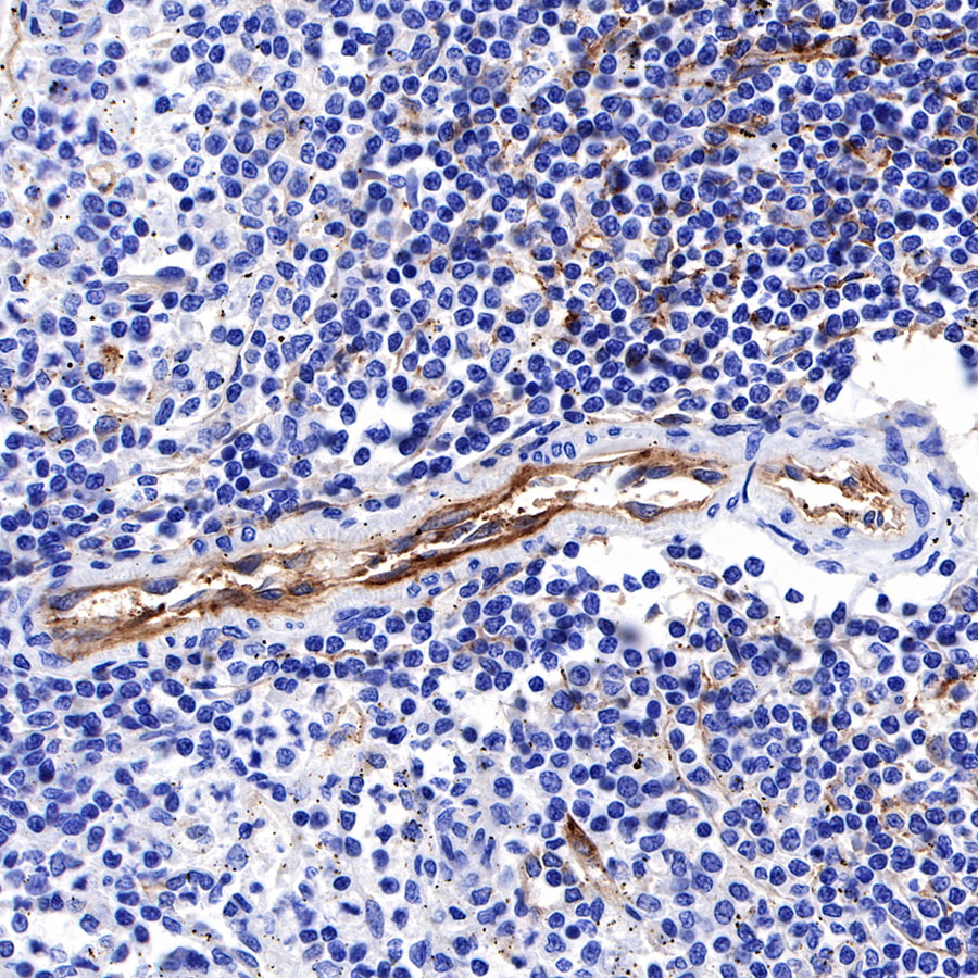
IHC shows positive staining in paraffin-embedded human spleen. Anti-Thrombomodulin antibody was used at 1/1000 dilution, followed by a HRP Polymer for Mouse & Rabbit IgG (ready to use). Counterstained with hematoxylin. Heat mediated antigen retrieval with Tris/EDTA buffer pH9.0 was performed before commencing with IHC staining protocol.
IHC shows positive staining in paraffin-embedded human stomach. Anti-Thrombomodulin antibody was used at 1/1000 dilution, followed by a HRP Polymer for Mouse & Rabbit IgG (ready to use). Counterstained with hematoxylin. Heat mediated antigen retrieval with Tris/EDTA buffer pH9.0 was performed before commencing with IHC staining protocol.
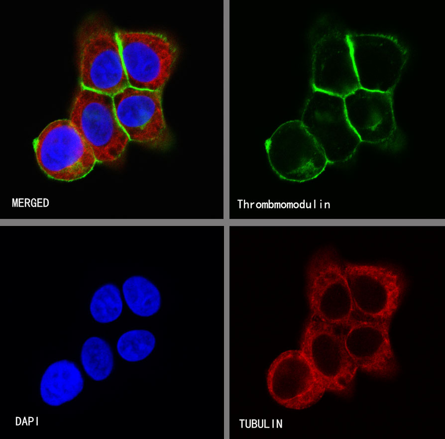
ICC shows positive staining in A431 cells. Anti-Thrombomodulin antibody was used at 1/250 dilution (Green) and incubated overnight at 4°C. Goat polyclonal Antibody to Rabbit IgG - H&L (Alexa Fluor® 488) was used as secondary antibody at 1/1000 dilution. The cells were fixed with 100% ice-cold methanoland permeabilized with 0.1% PBS-Triton X-100. Nuclei were counterstained with DAPI (Blue).Counterstain with tubulin (Red).
 您现在的位置: