

PBS, 40% Glycerol, 0.05% BSA, 0.03% Proclin 300
12 months from date of receipt / reconstitution, -20 °C as supplied
| 应用 | 稀释度 |
|---|---|
| WB | 1:4000-1:20000 |
| IHC-P | 1:1000 |
Tubulin is the major constituent of microtubules, a cylinder consisting of laterally associated linear protofilaments composed of alpha- and beta-tubulin heterodimers. Tubulin α- and β-subunits have molecular weights of ~ 50 kDa and are 36%–42% identical and 63% homologous. Both tubulin subunits bind guanine nucleotides. The binding to α-tubulin at the N-site is nonexchangeable, while the binding to β-tubulin at the E-site is exchangeable. Nucleotide in microtubules does not exchange with the solution, except for terminal subunits at microtubule ends.
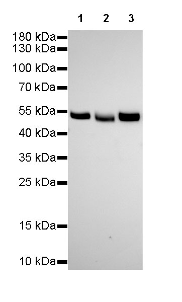
WB result of α-tubulin Rabbit mAb
Primary antibody: α-tubulin Rabbit mAb at 1/4000 dilution
Lane 1: HeLa whole cell lysate 20 µg
Lane 2: HepG2 whole cell lysate 20 µg
Lane 3: Jurkat whole cell lysate 20 µg
Secondary antibody: Goat Anti-Rabbit IgG, (H+L), HRP conjugated at 1/10000 dilution
Predicted MW: 52 kDa
Observed MW: 52 kDa
Exposure time: 150s
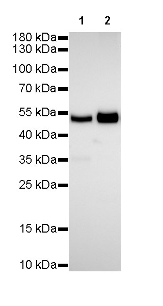
WB result of α-tubulin Rabbit mAb
Primary antibody: α-tubulin Rabbit mAb at 1/4000 dilution
Lane 1: NIH/3T3 whole cell lysate 20 µg
Lane 2: mouse brain lysate 5 µg
Secondary antibody: Goat Anti-Rabbit IgG, (H+L), HRP conjugated at 1/10000 dilution
Predicted MW: 52 kDa
Observed MW: 52 kDa
Exposure time: 150s
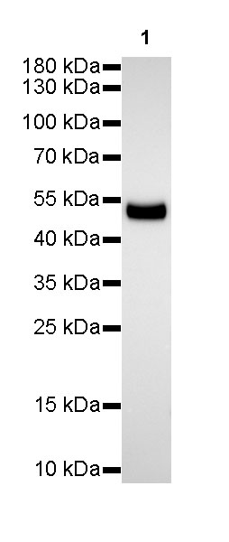
WB result of α-tubulin Rabbit mAb
Primary antibody: α-tubulin Rabbit mAb at 1/4000 dilution
Lane 1: rat brain lysate 5 µg
Secondary antibody: Goat Anti-Rabbit IgG, (H+L), HRP conjugated at 1/10000 dilution
Predicted MW: 52 kDa
Observed MW: 52 kDa
Exposure time: 60s
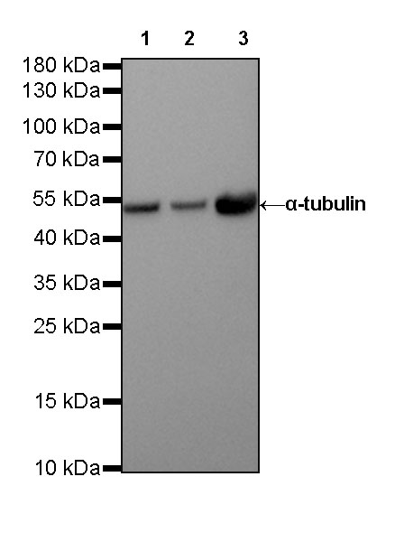
WB result of α-tubulin Rabbit mAb
Primary antibody: α-tubulin Rabbit mAb at 1/20000 dilution
Lane 1: HeLa whole cell lysate 20 µg
Lane 2: NIH/3T3 whole cell lysate 20 µg
Lane 3: rat brain lysate 20 µg
Secondary antibody: Goat Anti-Rabbit IgG, (H+L), HRP conjugated at 1/10000 dilution
Predicted MW: 52 kDa
Observed MW: 52 kDa
Exposure time: 90 s
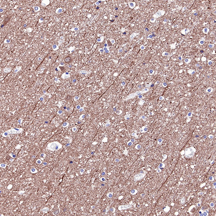
IHC shows positive staining in paraffin-embedded human cerebral cortex. Anti-α-tubulin antibody was used at 1/1000 dilution, followed by a HRP Polymer for Mouse & Rabbit IgG (ready to use). Counterstained with hematoxylin. Heat mediated antigen retrieval with Tris/EDTA buffer pH9.0 was performed before commencing with IHC staining protocol.
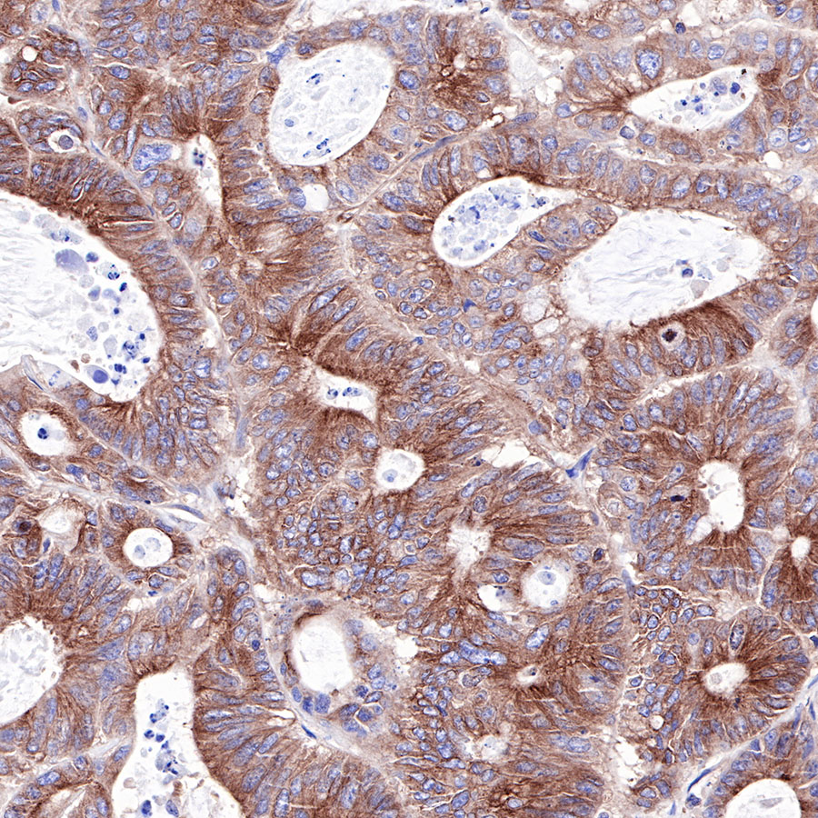
IHC shows positive staining in paraffin-embedded human colon cancer. Anti-α-tubulin antibody was used at 1/1000 dilution, followed by a HRP Polymer for Mouse & Rabbit IgG (ready to use). Counterstained with hematoxylin. Heat mediated antigen retrieval with Tris/EDTA buffer pH9.0 was performed before commencing with IHC staining protocol.
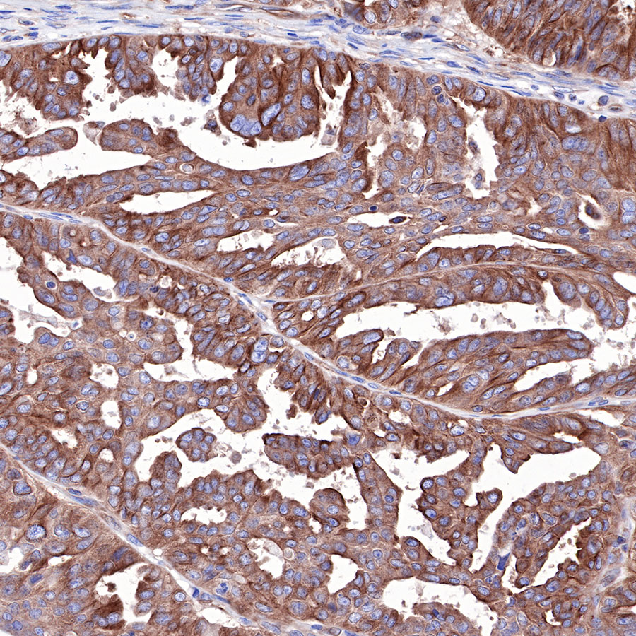
IHC shows positive staining in paraffin-embedded human ovarian cancer. Anti-α-tubulin antibody was used at 1/1000 dilution, followed by a HRP Polymer for Mouse & Rabbit IgG (ready to use). Counterstained with hematoxylin. Heat mediated antigen retrieval with Tris/EDTA buffer pH9.0 was performed before commencing with IHC staining protocol.
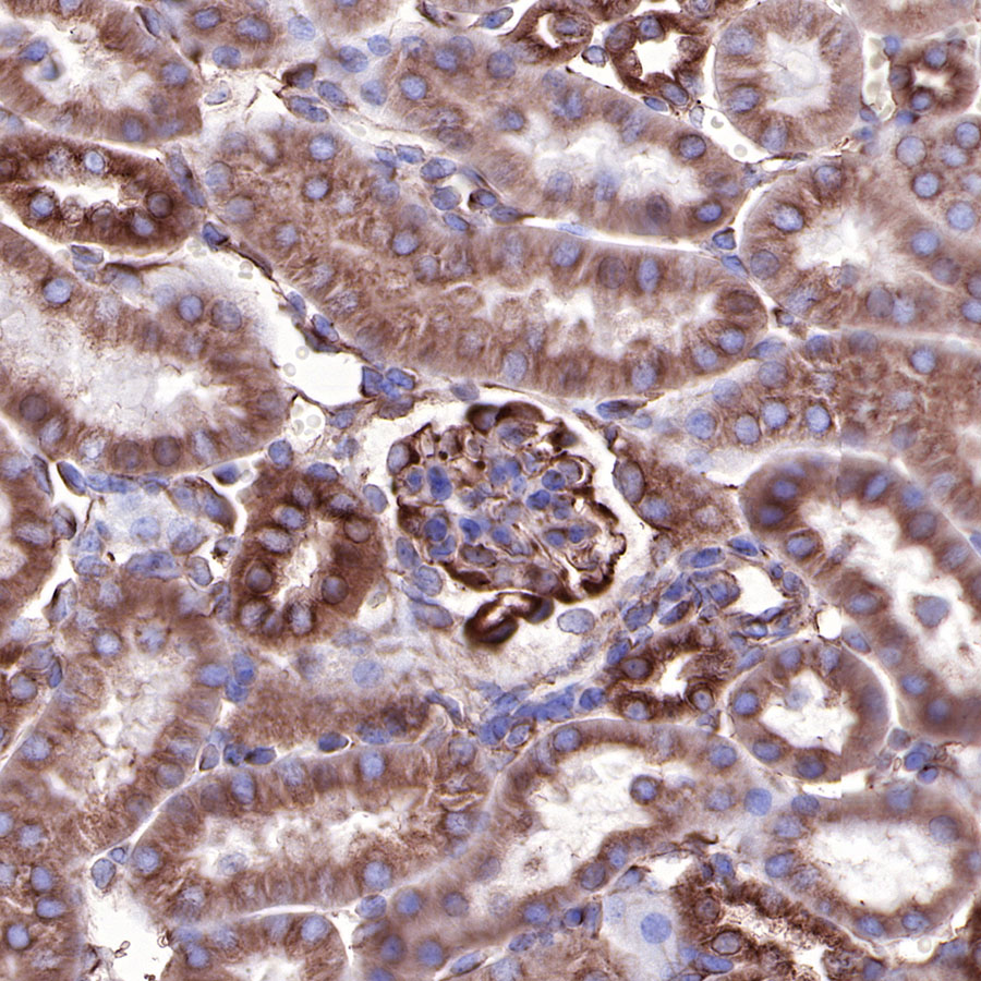
IHC shows positive staining in paraffin-embedded mouse kidney. Anti-α-tubulin antibody was used at 1/1000 dilution, followed by a HRP Polymer for Mouse & Rabbit IgG (ready to use). Counterstained with hematoxylin. Heat mediated antigen retrieval with Tris/EDTA buffer pH9.0 was performed before commencing with IHC staining protocol.
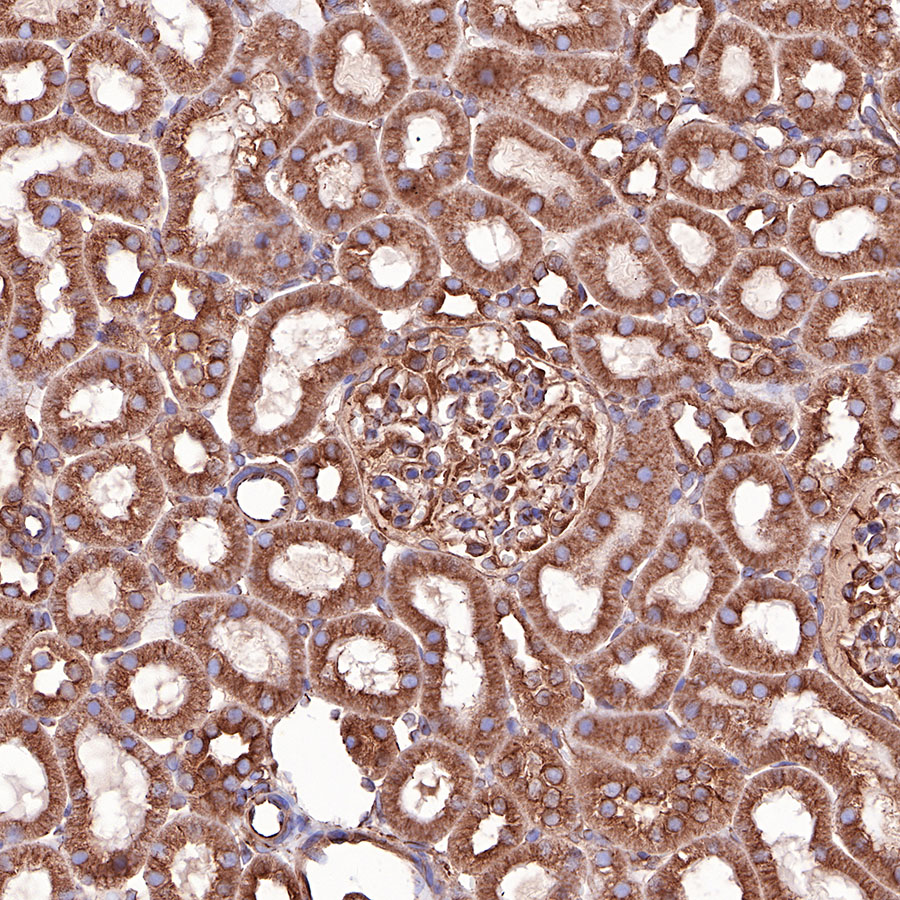
IHC shows positive staining in paraffin-embedded rat kidney. Anti-α-tubulin antibody was used at 1/1000 dilution, followed by a HRP Polymer for Mouse & Rabbit IgG (ready to use). Counterstained with hematoxylin. Heat mediated antigen retrieval with Tris/EDTA buffer pH9.0 was performed before commencing with IHC staining protocol.
 您现在的位置: