

12 months from date of receipt / reconstitution, -20 °C as supplied
| 应用 | 稀释度 |
|---|---|
| WB | 1:500 |
| IHC-P | 1:1000 |
| IP | 1:25 |
| ICC | 1:50 |
| ICFCM | 1:250 |
Oct-4, a transcription factor also known as Oct-3, Oct-3/4, Otf3 or NF-A3, is encoded by the Pou5f1 gene (located on chromosome 6 in human and 17 in mouse) and belongs to the POU (Pit, Oct, Unc) family of DNA binding-proteins. These proteins regulate the expression of target genes by binding to the octamer motif ATGCAAAT within their promoter or enhancer regions. Oct4, whose expression is associated with pluripotent properties of stem cells, is an essential factor controlling early stages of mammalian embryogenesis.
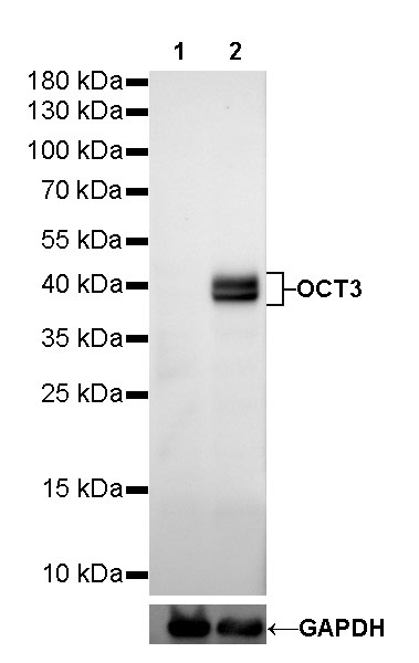
WB result of OCT3/4 Rabbit mAb
Primary antibody: OCT3/4 Rabbit mAb at 1/500 dilution
Lane 1: HeLa whole cell lysate 20 µg
Lane 2: NCCIT whole cell lysate 20 µg
Negative control: HeLa whole cell lysate
Secondary antibody: Goat Anti-Rabbit IgG, (H+L), HRP conjugated at 1/10000 dilution
Predicted MW: 45 kDa
Observed MW: 40 kDa
Exposure time: 60s
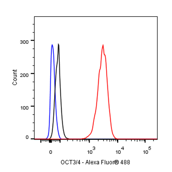
Flow cytometric analysis of 4% PFA fixed 90% methanol permeabilized NCCIT (Human pluripotent embryonic carcinoma epithelial cell) cells labelling Amyloid Precursor Protein antibody at 1/250 dilution (0.1 μg)/ (Red) compared with a Rabbit monoclonal IgG (Black) isotype control and an unlabelled control (cells without incubation with primary antibody and secondary antibody) (Blue). Goat Anti - Rabbit IgG Alexa Fluor® 488 was used as the secondary antibody.

OCT3/4 Rabbit mAb at 1/25 dilution (1µg) immunoprecipitating OCT3/4 in 0.4mg NCCIT whole cell lysate.
Western blot was performed on the immunoprecipitate using OCT3/4 Rabbit mAb at 1/1000 dilution.
Secondary antibody (HRP) for IP was used at 1/400 dilution.
Lane 1 : NCCIT whole cell lysate 10µg(input)
Lane 2 : OCT3/4 Rabbit mAb IP in NCCIT whole cell lysate
Lane 3 : Rabbit monoclonal IgG IP in NCCIT whole cell lysate
Predicted MW: 45 kDa
Observed MW: 40 kDa
Exposure time: 20s
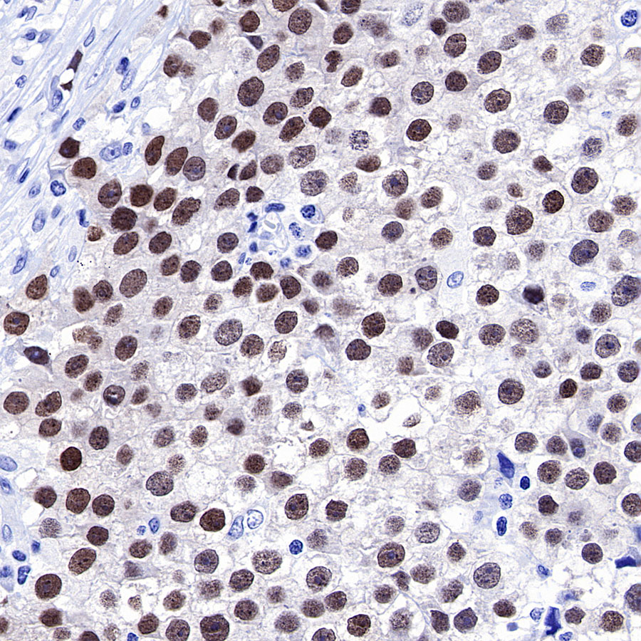
IHC shows positive staining in paraffin-embedded human seminoma. Anti-OCT3/4 antibody was used at 1/1000 dilution, followed by a HRP Polymer for Mouse & Rabbit IgG (ready to use). Counterstained with hematoxylin. Heat mediated antigen retrieval with Tris/EDTA buffer pH9.0 was performed before commencing with IHC staining protocol.
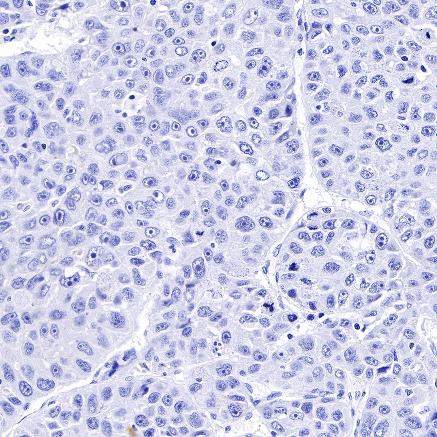
Negative control: IHC shows negative staining in paraffin-embedded human hepatocellular carcinoma. Anti-OCT3/4 antibody was used at 1/1000 dilution, followed by a HRP Polymer for Mouse & Rabbit IgG (ready to use). Counterstained with hematoxylin. Heat mediated antigen retrieval with Tris/EDTA buffer pH9.0 was performed before commencing with IHC staining protocol.
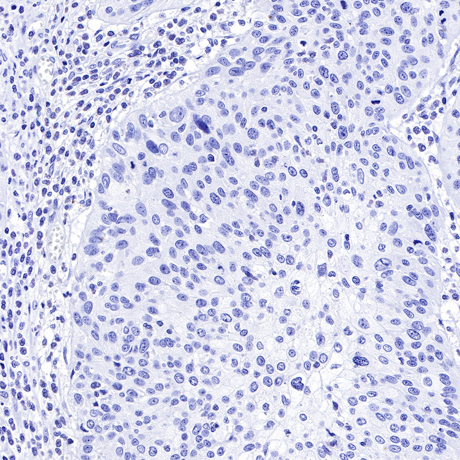
Negative control: IHC shows negative staining in paraffin-embedded human cervical carcinoma. Anti-OCT3/4 antibody was used at 1/1000 dilution, followed by a HRP Polymer for Mouse & Rabbit IgG (ready to use). Counterstained with hematoxylin. Heat mediated antigen retrieval with Tris/EDTA buffer pH9.0 was performed before commencing with IHC staining protocol.
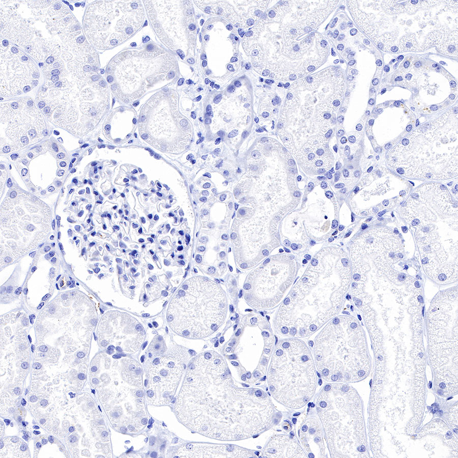
Negative control: IHC shows negative staining in paraffin-embedded human kidney. Anti-OCT3/4 antibody was used at 1/1000 dilution, followed by a HRP Polymer for Mouse & Rabbit IgG (ready to use). Counterstained with hematoxylin. Heat mediated antigen retrieval with Tris/EDTA buffer pH9.0 was performed before commencing with IHC staining protocol.
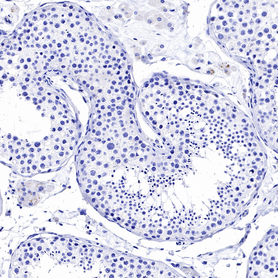
Negative control: IHC shows negative staining in paraffin-embedded human testis. Anti-OCT3/4 antibody was used at 1/1000 dilution, followed by a HRP Polymer for Mouse & Rabbit IgG (ready to use). Counterstained with hematoxylin. Heat mediated antigen retrieval with Tris/EDTA buffer pH9.0 was performed before commencing with IHC staining protocol.
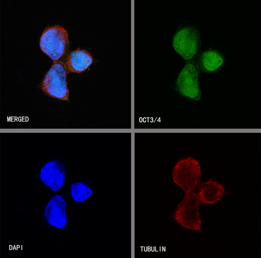
ICC shows positive staining in NCCIT cells. Anti-OCT3/4 antibody was used at 1/50 dilution (Green) and incubated overnight at 4°C. Goat polyclonal Antibody to Rabbit IgG - H&L (Alexa Fluor® 488) was used as secondary antibody at 1/1000 dilution. The cells were fixed with 100% ice-cold methanol and permeabilized with 0.1% PBS-Triton X-100. Nuclei were counterstained with DAPI (Blue). Counterstain with tubulin (red).
 您现在的位置: