
PBS, 40% Glycerol, 0.05% BSA, 0.03% Proclin 300
12 months from date of receipt / reconstitution, -20 °C as supplied
| 应用 | 稀释度 | 推荐种属 |
|---|---|---|
| WB | 1:1000 | Hu, Ms, Rt |
| IHC-P | 1:250 | Hu |
| ICC | 1:500 | Hu |
| IF | 1:500 | Hu |
The Wilms Tumor Protein (WT1) is a transcription factor that plays a crucial role in the development and differentiation of various tissues, particularly in the kidneys and gonads. It is encoded by the WT1 gene and is essential for normal nephrogenesis, ensuring the proper formation of the glomeruli and other renal structures. Abnormal expression or mutations in the WT1 gene are associated with Wilms tumor, a pediatric kidney cancer, as well as other developmental disorders such as Denys-Drash syndrome and Frasier syndrome. In addition to its role in development, WT1 is also involved in the regulation of cell proliferation, apoptosis, and the maintenance of mesenchymal stem cells. Its expression is tightly regulated and has been studied extensively as a potential biomarker and therapeutic target in cancer research.
WB result of Wilms Tumor Protein (WT1) Recombinant Rabbit mAb
Primary antibody: Wilms Tumor Protein (WT1) Recombinant Rabbit mAb at 1/1000 dilution
Lane 1: DU 145 whole cell lysate 20 µg
Lane 2: K562 whole cell lysate 20 µg
Lane 3: OVCAR-3 whole cell lysate 20 µg
Negative control: DU 145 whole cell lysate
Secondary antibody: Goat Anti-rabbit IgG, (H+L), HRP conjugated at 1/10000 dilution
Predicted MW: 50 kDa
Observed MW: 55 kDa
WB result of Wilms Tumor Protein (WT1) Recombinant Rabbit mAb
Primary antibody: Wilms Tumor Protein (WT1) Recombinant Rabbit mAb at 1/1000 dilution
Lane 1: mouse testis lysate 20 µg
Secondary antibody: Goat Anti-rabbit IgG, (H+L), HRP conjugated at 1/10000 dilution
Predicted MW: 50 kDa
Observed MW: 53 kDa
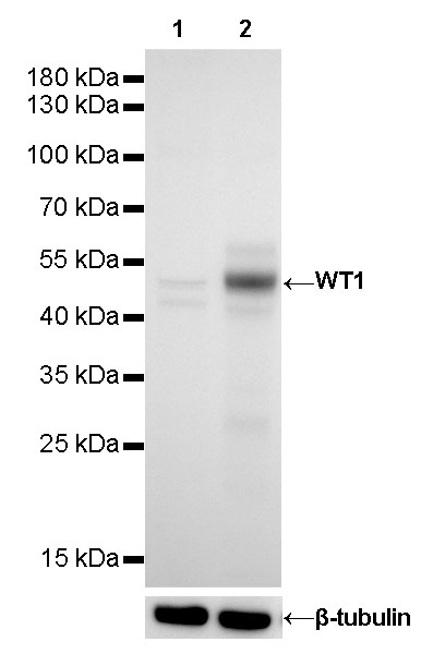
WB result of Wilms Tumor Protein (WT1) Recombinant Rabbit mAb
Primary antibody: Wilms Tumor Protein (WT1) Recombinant Rabbit mAb at 1/1000 dilution
Lane 1: rat testis lysate 20 µg
Secondary antibody: Goat Anti-rabbit IgG, (H+L), HRP conjugated at 1/10000 dilution
Predicted MW: 50 kDa
Observed MW: 53 kDa
IHC shows positive staining in paraffin-embedded Wilms tumor. Anti-Wilms Tumor Protein (WT1) antibody was used at 1/250 dilution, followed by a HRP Polymer for Mouse & Rabbit IgG (ready to use). Counterstained with hematoxylin. Heat mediated antigen retrieval with Tris/EDTA buffer pH9.0 was performed before commencing with IHC staining protocol.
IHC shows positive staining in paraffin-embedded human kidney. Anti-Wilms Tumor Protein (WT1) antibody was used at 1/250 dilution, followed by a HRP Polymer for Mouse & Rabbit IgG (ready to use). Counterstained with hematoxylin. Heat mediated antigen retrieval with Tris/EDTA buffer pH9.0 was performed before commencing with IHC staining protocol.
IHC shows positive staining in paraffin-embedded human ovarian cancer. Anti-Wilms Tumor Protein (WT1) antibody was used at 1/250 dilution, followed by a HRP Polymer for Mouse & Rabbit IgG (ready to use). Counterstained with hematoxylin. Heat mediated antigen retrieval with Tris/EDTA buffer pH9.0 was performed before commencing with IHC staining protocol.
ICC shows positive staining in OVCAR-3 cells. Anti- Wilms Tumor Protein (WT1) antibody was used at 1/500 dilution (Green) and incubated overnight at 4°C. Goat polyclonal Antibody to Rabbit IgG - H&L (Alexa Fluor® 488) was used as secondary antibody at 1/1000 dilution. The cells were fixed with 4% PFA and permeabilized with 0.1% PBS-Triton X-100. Nuclei were counterstained with DAPI (Blue). Counterstain with tubulin (Red).
IF shows positive staining in paraffin-embedded human ovarian cancer. Anti- Wilms Tumor Protein (WT1) antibody was used at 1/500 dilution (Green) and incubated overnight at 4°C. Goat polyclonal Antibody to Rabbit IgG - H&L (Alexa Fluor® 488) was used as secondary antibody at 1/1000 dilution. Counterstained with DAPI (Blue). Heat mediated antigen retrieval with EDTA buffer pH9.0 was performed before commencing with IF staining protocol.
IF shows positive staining in paraffin-embedded human lung squamous carcinoma. Anti- Wilms Tumor Protein (WT1) antibody was used at 1/500 dilution (Green) and incubated overnight at 4°C. Goat polyclonal Antibody to Rabbit IgG - H&L (Alexa Fluor® 488) was used as secondary antibody at 1/1000 dilution. Counterstained with DAPI (Blue). Heat mediated antigen retrieval with EDTA buffer pH9.0 was performed before commencing with IF staining protocol.
Expression of Wilms Tumor Protein (WT1) in tumor tissue.
Expression of Wilms Tumor Protein (WT1) in human tissue.
 您现在的位置: