
12 months from date of receipt / reconstitution, -20 °C as supplied
| 应用 | 稀释度 |
|---|---|
| IHC-P | 1:1000 |
| FC | 1:250 |
| ICC | 1:250 |
| WB | 1:500 |
Paired box gene 8, also known as PAX8, is a protein which in humans is encoded by the PAX8 gene. This gene is a member of the paired box (PAX) family of transcription factors. Members of this gene family typically encode proteins which contain a paired box domain, an octapeptide, and a paired-type homeodomain. The PAX gene family has an important role in the formation of tissues and organs during embryonic development and maintaining the normal function of some cells after birth. PAX8 (and PAX2) is one of the important regulators of urogenital system morphogenesis.

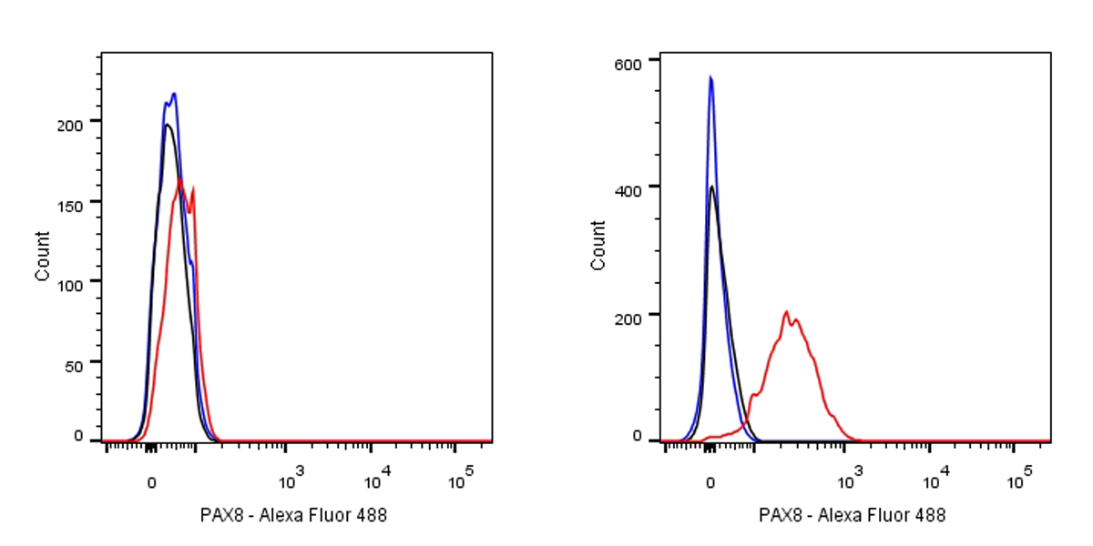
Flow cytometric analysis of HeLa (left) / SK-OV-3 (right) cells labelling PAX8 antibody at 1/250 dilution (0.1ug)/ (red) compared with a Rabbit monoclonal IgG (Black) isotype control and an unlabelled control (cells without incubation with primary antibody and secondary antibody) (Blue).
Goat Anti-Rabbit IgG Alexa Fluor® 488 at 1/1000 dilution was used as the secondary antibody.
Negative control: HeLa
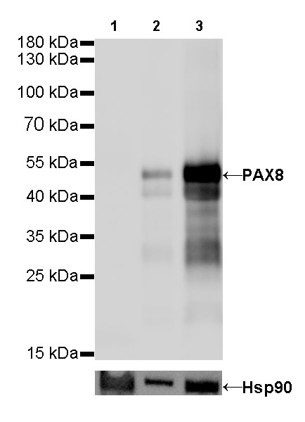
WB result of PAX8 Rabbit mAb
Primary antibody: PAX8 Rabbit mAb at 1/500 dilution
Lane 1: Hela whole cell lysate 20 µg
Lane 2: SK-OV-3 whole cell lysate 20 µg
Lane 3: OVCAR-3 whole cell lysate 20 µg
Negative control: Hela whole cell lysate
Secondary antibody: Goat Anti-Rabbit IgG, (H+L), HRP conjugated at 1/10000 dilution
Predicted MW: 48 kDa
Observed MW: 48 kDa
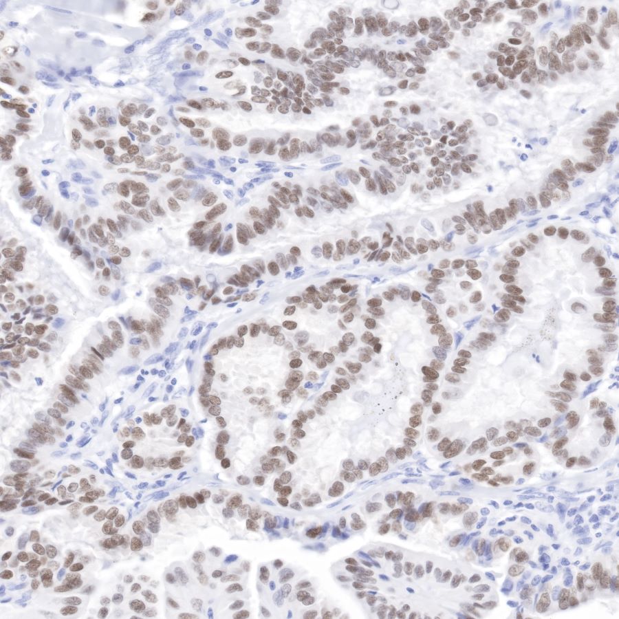
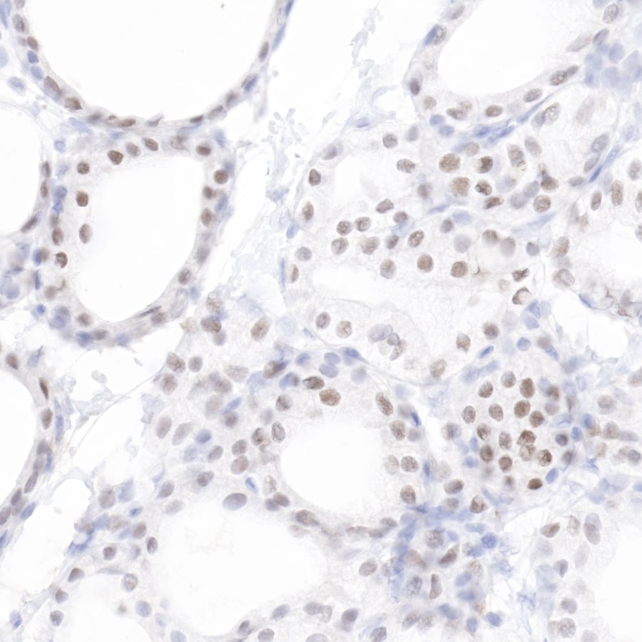
IHC shows positive staining in paraffin-embedded human thyroid cancer.
Anti-PAX8 antibody was used at 1/1000 dilution, followed by a Goat Anti-Rabbit IgG H&L (HRP) ready to use. Counterstained with hematoxylin.
Heat mediated antigen retrieval with Tris/EDTA buffer pH9.0 was performed before commencing with IHC staining protocol.
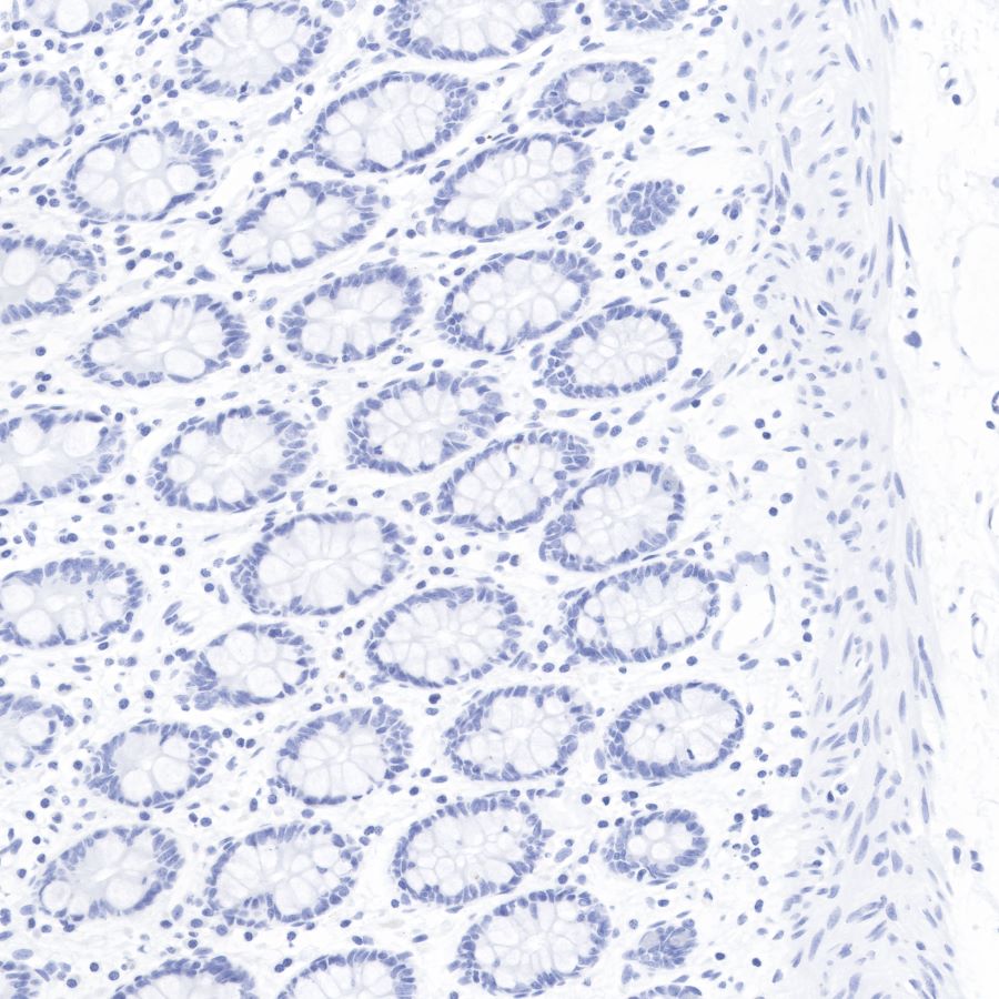
Negative control: IHC shows negative staining in paraffin-embedded human colon.
Anti-PAX8 antibody was used at 1/1000 dilution, followed by a Goat Anti-Rabbit IgG H&L (HRP) ready to use. Counterstained with hematoxylin.
Heat mediated antigen retrieval with Tris/EDTA buffer pH9.0 was performed before commencing with IHC staining protocol.
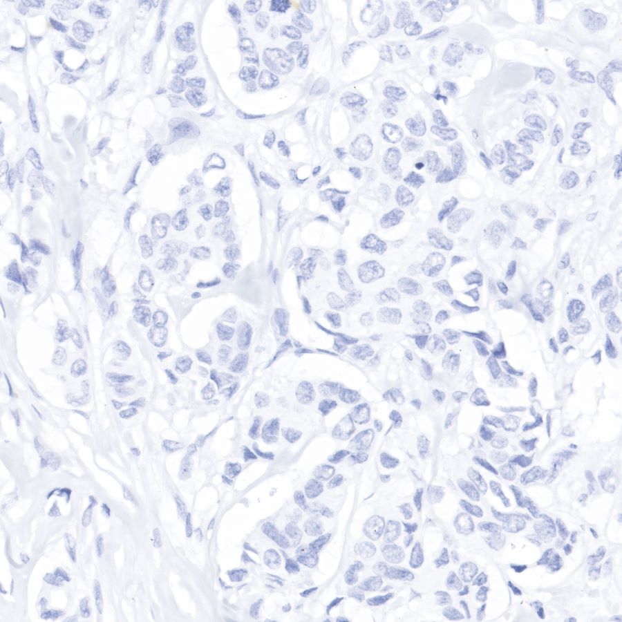
Negative control: IHC shows negative staining in paraffin-embedded human breast cancer.
Anti-PAX8 antibody was used at 1/1000 dilution, followed by a Goat Anti-Rabbit IgG H&L (HRP) ready to use.
Counterstained with hematoxylin.
Heat mediated antigen retrieval with Tris/EDTA buffer pH9.0 was performed before commencing with IHC staining protocol.

IHC shows positive staining in paraffin-embedded mouse thyroid.
Anti-PAX8 antibody was used at 1/1000 dilution, followed by a Goat Anti-Rabbit IgG H&L (HRP) ready to use.
Counterstained with hematoxylin.
Heat mediated antigen retrieval with Tris/EDTA buffer pH9.0 was performed before commencing with IHC staining protocol.
IHC shows positive staining in paraffin-embedded rat thyroid. Anti-PAX8 antibody was used at 1/1000 dilution, followed by a Goat Anti-Rabbit IgG H&L (HRP) ready to use.
Counterstained with hematoxylin.
Heat mediated antigen retrieval with Tris/EDTA buffer pH9.0 was performed before commencing with IHC staining protocol.PA
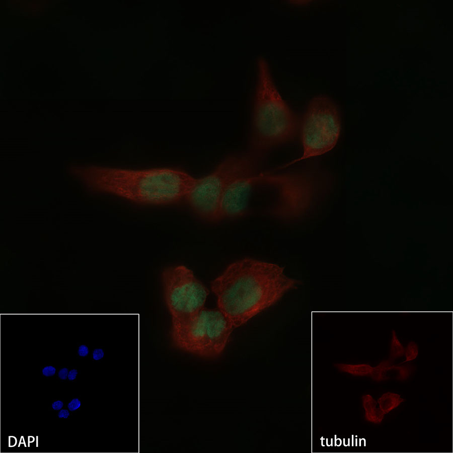
ICC shows nuclear staining in SK-OV-3 cells.
Anti-PAX8 antibody was used at 1/250 dilution and incubated overnight at 4°C. Goat polyclonal Antibody to Rabbit IgG - H&L (Alexa Fluor® 488) was used as secondary antibody at 1/1000 dilution.
The cells were fixed with 4% PFA and permeabilized with 0.1% PBS-Triton X-100.
Nuclei were counterstained with DAPI.
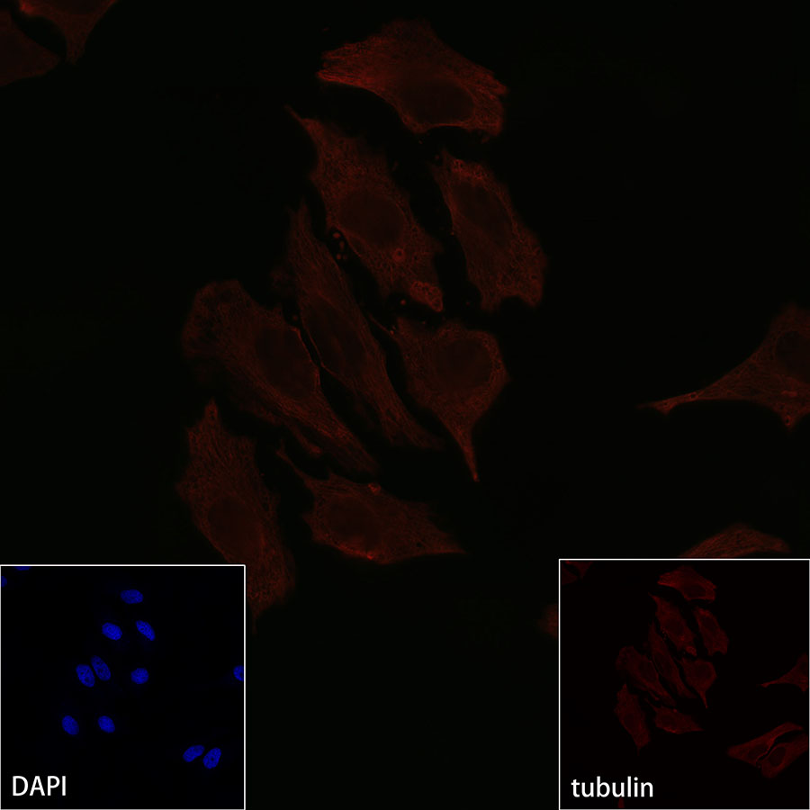
Negative control: ICC shows negative staining in HeLa cells. Anti-PAX8 antibody was used at 1/250 dilution and incubated overnight at 4°C.
Goat polyclonal Antibody to Rabbit IgG - H&L (Alexa Fluor® 488) was used as secondary antibody at 1/1000 dilution.
The cells were fixed with 4% PFA and permeabilized with 0.1% PBS-Triton X-100. Nuclei were counterstained with DAPI.
 您现在的位置: