

PBS, 40% Glycerol, 0.05%BSA, 0.03% Proclin 300
12 months from date of receipt / reconstitution, -20 °C as supplied
| 应用 | 稀释度 |
|---|---|
| FCM | 1:250 |
| WB | 1:10000-1:20000 |
| IHC-P | 1:1000 |
| ICC | 1:250 |
Desmin is a protein that in humans is encoded by the DES gene. Desmin is a subunit of intermediate filaments in cardiac muscle, skeletal muscle and smooth muscle tissue. In cardiac muscle, desmin is present in Z-discs and intercalated discs and regulates sarcomere architecture. Desmin-related myofibrillar myopathy (DRM or desminopathy) is a subgroup of the myofibrillar myopathy diseases and is the result of a mutation in the gene that codes for desmin which by changing the protein structure prevents it from forming protein filaments, and rather, forms aggregates of desmin and other proteins throughout the cell. Desmin has been evaluated for role in assessing the depth of invasion of urothelial carcinoma in TURBT specimens.
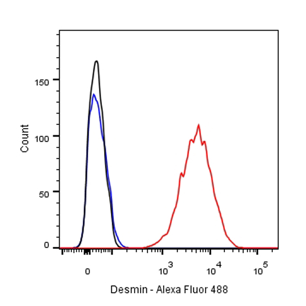
Flow cytometric analysis of C2C12 cells labelling Desmin antibody at 1/250 dilution/ (red) compared with a Rabbit monoclonal IgG (Black) isotype control and an unlabelled control (cells without incubation with primary antibody and secondary antibody) (Blue). Goat Anti-Rabbit IgG Alexa Fluor® 488 at 1/1000 dilution was used as the secondary antibody.
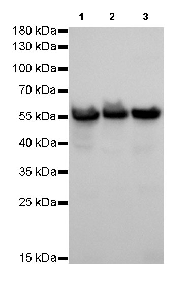
WB result of Desmin Rabbit mAb
Primary antibody: Desmin Rabbit mAb at 1/10000 dilution
Lane 1: C2C12 whole cell lysate 20 µg
Lane 2: mouse skeletal muscle lysate 20 µg
Lane 3: mouse heart lysate 20 µg
Secondary antibody: Goat Anti-Rabbit IgG, (H+L), HRP conjugated at 1/10000 dilution
Predicted MW: 53 kDa
Observed MW: 53 kDa
Exposure time: 0.5s
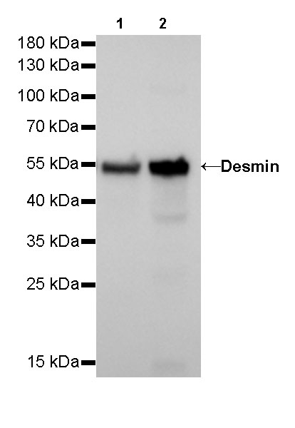
WB result of Desmin Rabbit mAb
Primary antibody: Desmin Rabbit mAb at 1/10000 dilution
Lane 1: rat skeletal muscle lysate 20 µg
Lane 2: rat heart lysate 20 µg
Secondary antibody: Goat Anti-Rabbit IgG, (H+L), HRP conjugated at 1/10000 dilution
Predicted MW: 53 kDa
Observed MW: 53 kDa
Exposure time: 0.5s
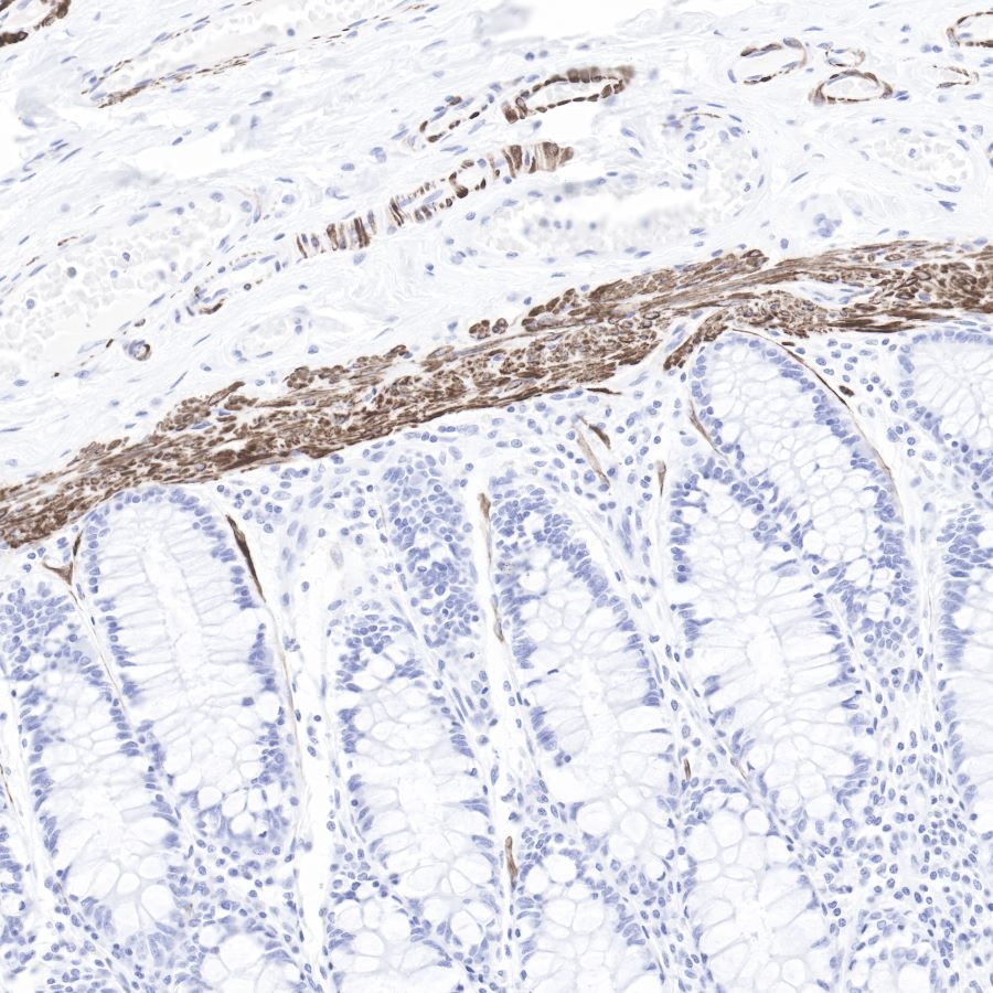
IHC shows positive staining in paraffin-embedded human colon.
Anti-Desmin antibody was used at 1/1000 dilution, followed by a Goat Anti-Rabbit IgG H&L (HRP) ready to use.
Counterstained with hematoxylin.
Heat mediated antigen retrieval with Tris/EDTA buffer pH9.0 was performed before commencing with IHC staining protocol.
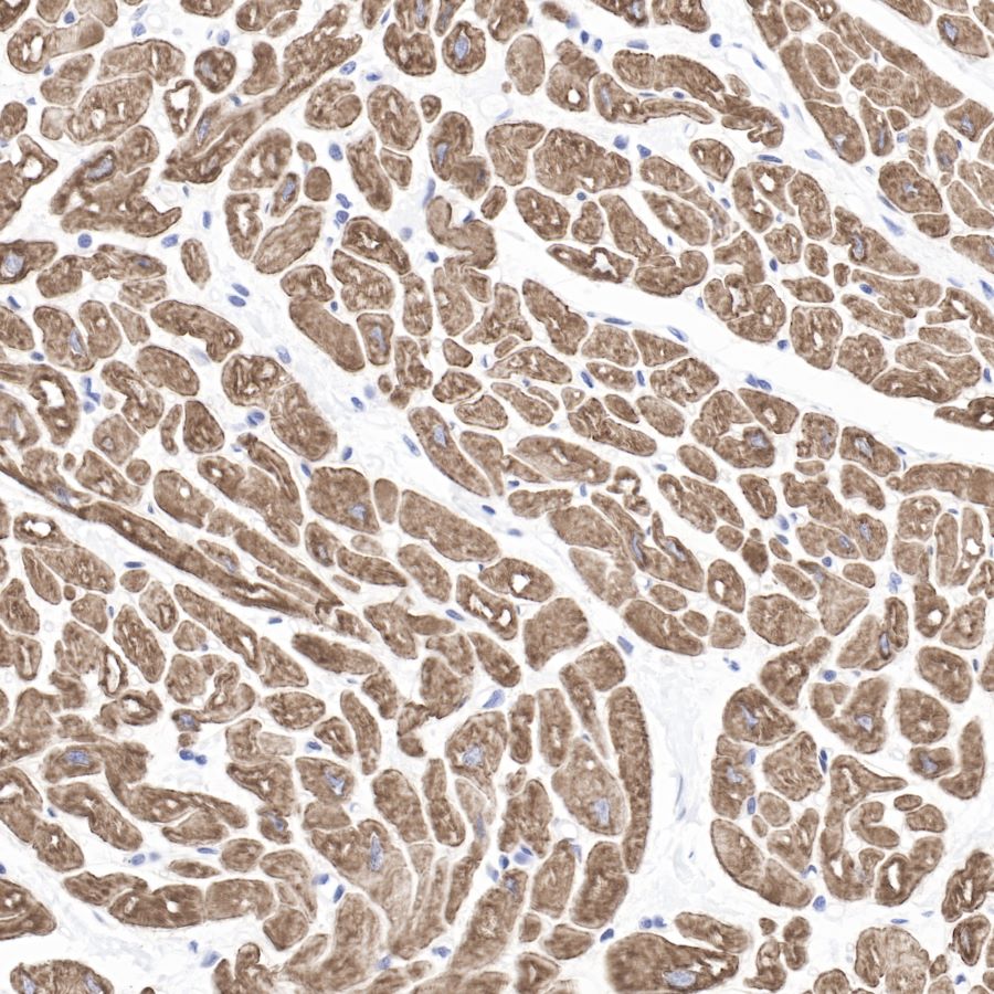
IHC shows positive staining in paraffin-embedded human cardiac muscle.
Anti-Desmin antibody was used at 1/1000 dilution, followed by a Goat Anti-Rabbit IgG H&L (HRP) ready to use.
Counterstained with hematoxylin.
Heat mediated antigen retrieval with Tris/EDTA buffer pH9.0 was performed before commencing with IHC staining protocol.
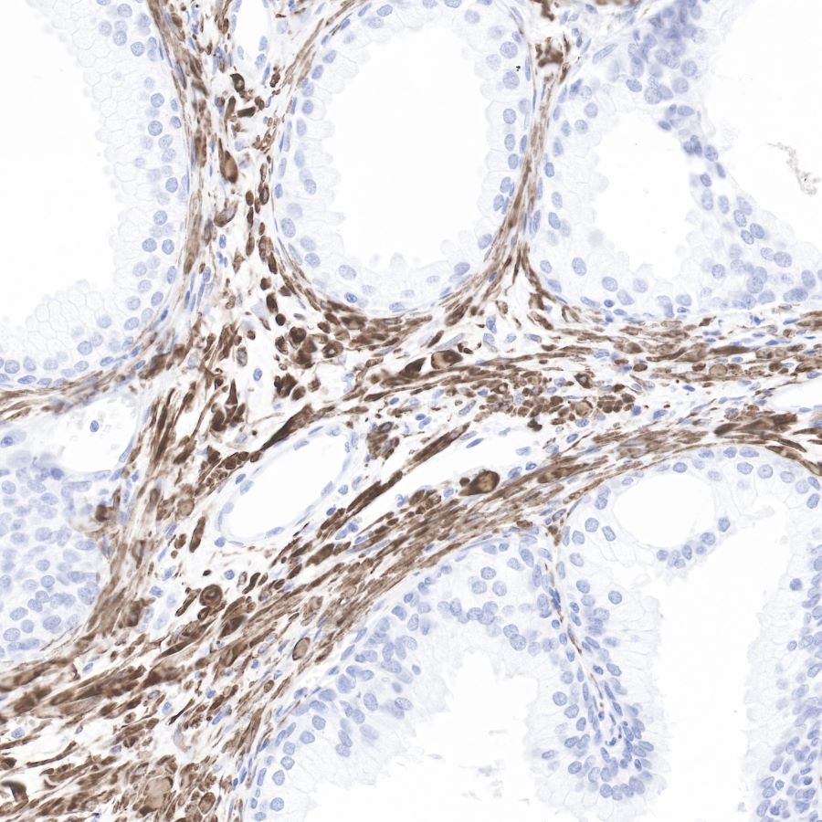
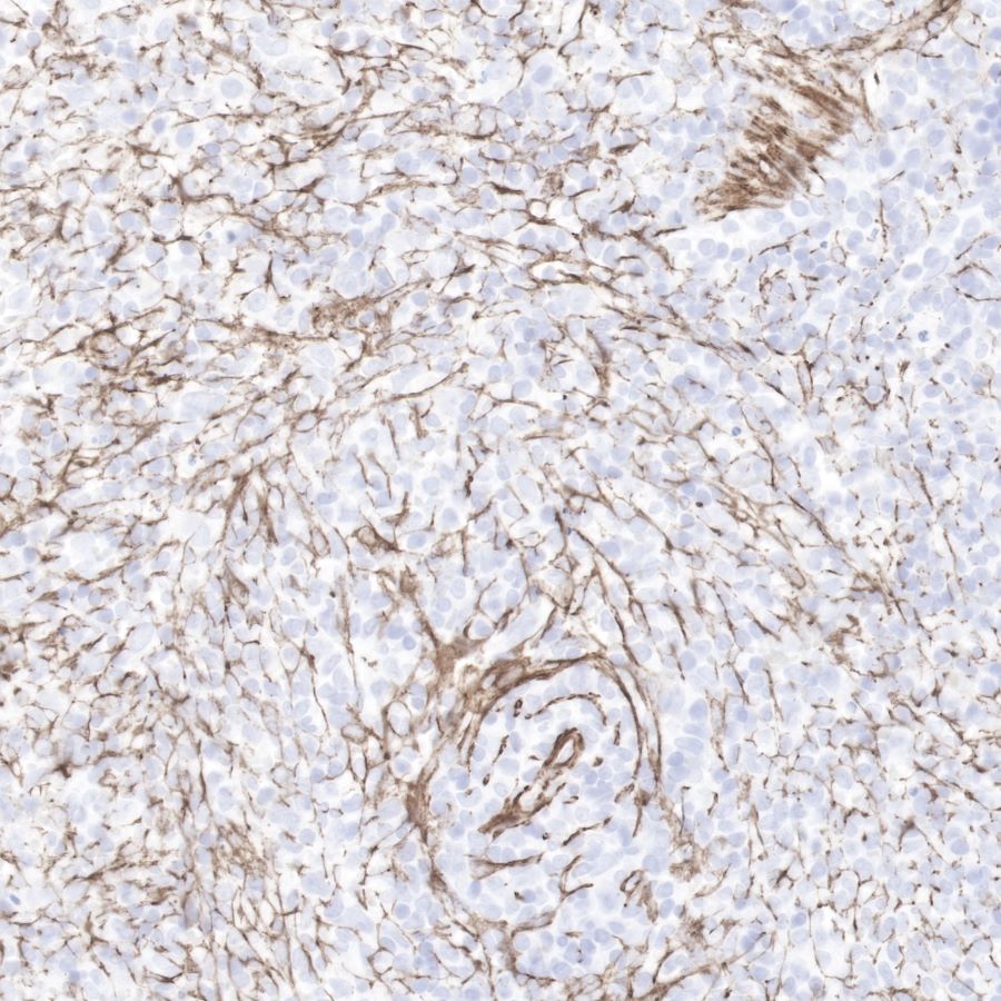
IHC shows positive staining in paraffin-embedded human prostate.
Anti-Desmin antibody was used at 1/1000 dilution, followed by a Goat Anti-Rabbit IgG H&L (HRP) ready to use.
Counterstained with hematoxylin.
Heat mediated antigen retrieval with Tris/EDTA buffer pH9.0 was performed before commencing with IHC staining protocol.
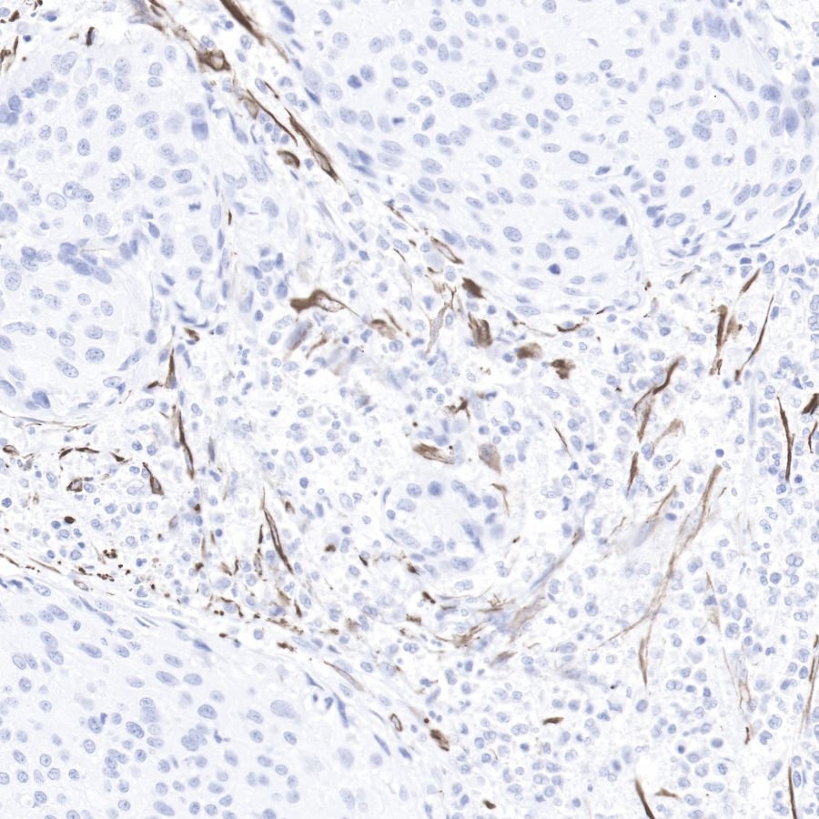
IHC shows positive staining in paraffin-embedded human cervix cancer.
Anti-Desmin antibody was used at 1/1000 dilution, followed by a Goat Anti-Rabbit IgG H&L (HRP) ready to use.
Counterstained with hematoxylin.
Heat mediated antigen retrieval with Tris/EDTA buffer pH9.0 was performed before commencing with IHC staining protocol.
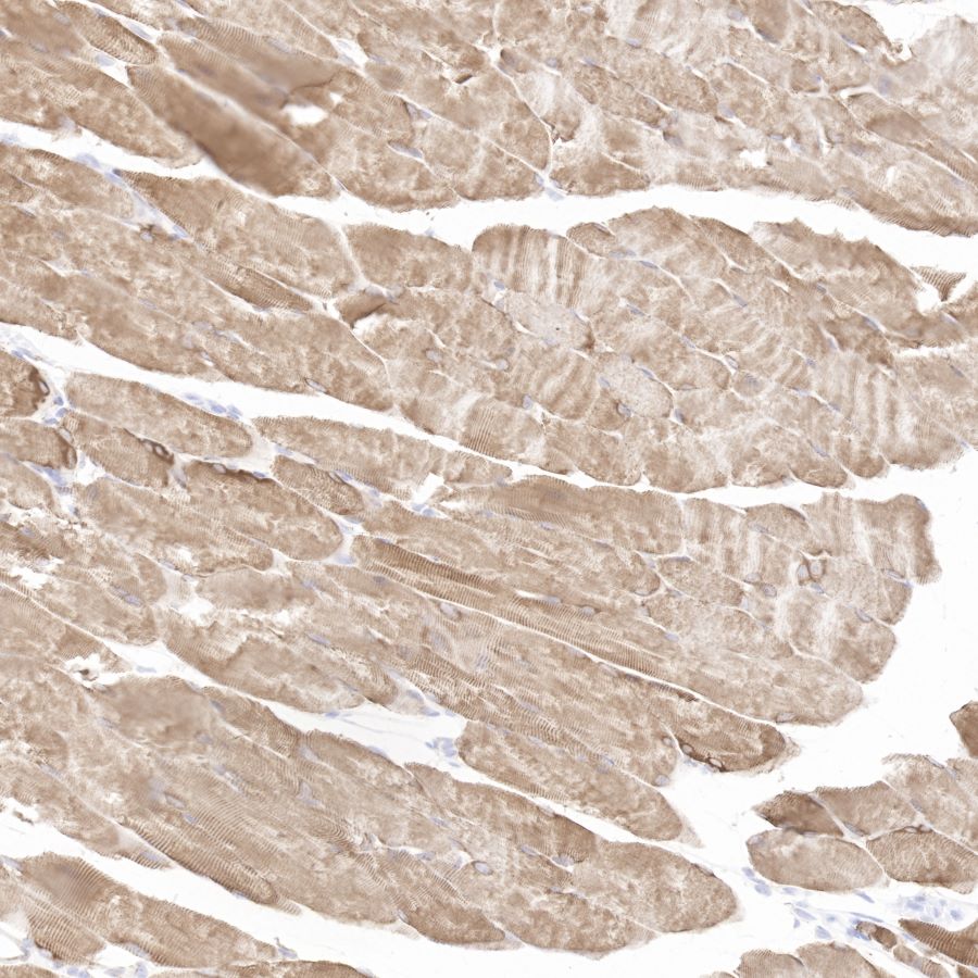
IHC shows positive staining in paraffin-embedded mouse skeletal muscle.
Anti-Desmin antibody was used at 1/1000 dilution, followed by a Goat Anti-Rabbit IgG H&L (HRP) ready to use.
Counterstained with hematoxylin.
Heat mediated antigen retrieval with Tris/EDTA buffer pH9.0 was performed before commencing with IHC staining protocol.
IHC shows positive staining in paraffin-embedded rat spleen.
Anti-Desmin antibody was used at 1/1000 dilution, followed by a Goat Anti-Rabbit IgG H&L (HRP) ready to use.
Counterstained with hematoxylin.
Heat mediated antigen retrieval with Tris/EDTA buffer pH9.0 was performed before commencing with IHC staining protocol.
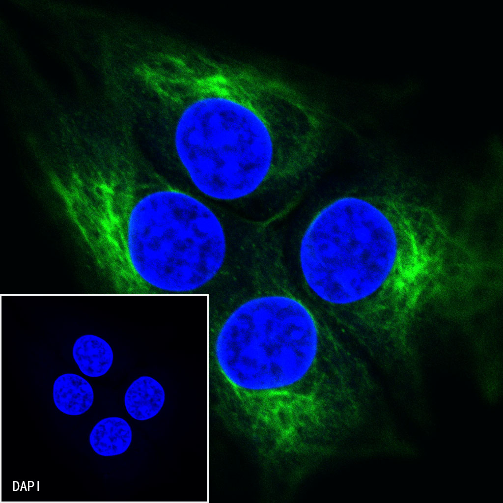
ICC shows positive staining in C2C12 cells. Anti-Desmin antibody was used at 1/250 dilution (Green) and incubated overnight at 4°C. Goat polyclonal Antibody to Rabbit IgG - H&L (Alexa Fluor® 488) was used as secondary antibody at 1/1000 dilution. The cells were fixed with 4%PFA and permeabilized with 0.1% PBS-Triton X-100. Nuclei were counterstained with DAPI (Blue).
 您现在的位置: